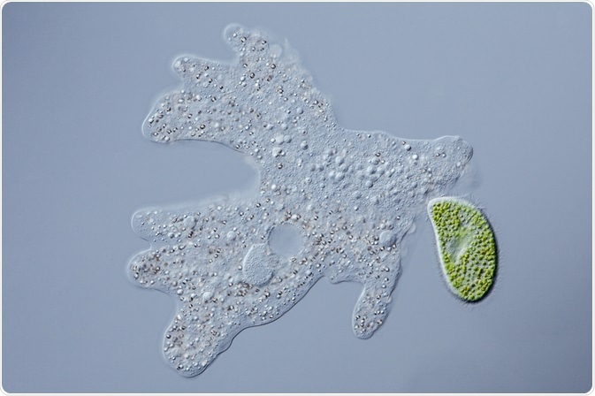
Reflection contrast microscopy is used to image cells with high resolution and improved sensitivity. As this technique can generate high definition images at high penetration depths, it is commonly used in immunocytochemistry.
 Lebendkulturen | Shutterstock
Method of reflection contrast microscopy
Reflection contrast microscopy can image small sections of cells by suppressing stray reflections. This is necessary when imaging biological samples, because biological objects tend to have reflected light intensities below 1% of incident light compared to 4% reflection intensity at a glass to air interface, which are common on microscopes.
The same amount of suppression achieved with this microscopy cannot be achieved using other microscopes. To suppress stray reflections, reflection contrast microscopy uses a monochromatic light source, typically 546 nm, while a polarizer and a lambda quarter wave plate lead to elliptical polarization and re-polarization of the anterior lens. Together with an aligned analyzer, exclusive reflections from the section reach the eyepiece.
The polarizers and wave plate used in reflection contrast microscopy account for the antiflex principle where object reflection light is turned 90° relative to the incident light, allowing for object imaging. The reflections achieved using reflection contrast microscopy are made with 45° epi-illumination. The conical, annular illumination merges the features of darkground illumination.
Reflection contrast microscopy can image incredibly thin sections, down to 5 nm, and a penetration depth of up to 30 µm. Because of this, 3D reconstructions can be created. Reflection contrast microscopy acts as a bridge between light microscopy and electron microscopy, and typically uses a fluorescence microscope stand with methodological adaptations to generate a reflection contrast microscopy image.
Applying reflection contrast microscopy to immunocytochemistry
Reflection contrast microscopy was originally developed for use in mineralogy and its applications in biology have only emerged very recently, including in situ hybridization and immunocytochemistry. This microscopy can be applied to living cells, which can be labelled using immunocytochemistry.
The first step is to fix the cells. It is important that the fixative is mild to maintain antigenicity, whereas strong fixatives are needed for morphological studies. In general, embedding and labelling molecules used in immunocytochemical localization studies can also be applied to reflection contrast microscopy studies.
There are two main methods to prepare a sample for reflection contrast microscopy: the immuno-incubation can be performed before or after the tissue is embedded.
Pre-embedding immunocytochemistry sees the tissue being sliced in 70–90 µm thick sections, which are then immunostained and embedded in plastic. Following this, they are cut into ultrathin sections. Common uses of pre-embedding immunocytochemistry are inter-cellular labeling of extracellular matrix in a tissue.
In post-embedding immunocytochemistry, the tissue is sliced immediately into ultrathin sections which are embedded and then stained. The type of embedding media used in post-embedding immunocytochemistry can affect the accessibility of antigenic sites for the antibodies, which may necessitate further etching steps, for example with Epon.
Post-embedding immunocytochemistry is often used to image the inside of a cell because the cell needs to be sectioned in ultrathin sections to achieve intracellular resolution.
Further Reading
Last Updated: Apr 1, 2019