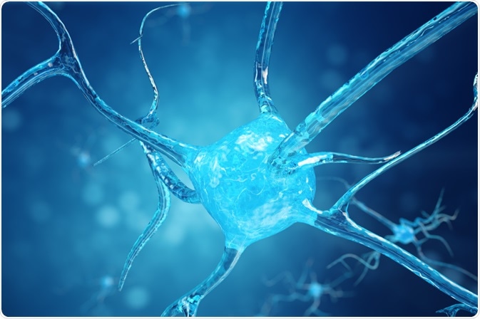
Connectomics is the study of the brain’s structural and functional connections between cells, which is visualized as a connectome.
 Rost9 | Shutterstock
The connectome is a map of all neural connections in a brain and connectomics is the mapping of these connections. Connectomics can provide insights about the brain and many incurable diseases that are associated with it.
The connectome
A connectome is also sometimes called a wiring diagram, wherein the brains axons and dendrites are analogous to wires and the neuron bodies to components. The molecular states related to learning at every synaptic connection is the synaptome, and changes in the nucleus of the neuron related to learning is referred to as the epigenome.
Connectomes tend to be animal-specific, such as one of the few completed connectomes of C. elegans, or specific to certain brain regions or subsystems, such as a hippocampal connectome.
The usefulness of connectomics is somewhat debated. Among popular arguments against it is that connectomics typically provides a static image. It can show which neurons have the possibility of interacting, but it does not show if they do, how much they do, and what the effect of that interaction is.
The key is integrating normal behaviors into the connectome, to see neuronal activity during normal states. However, a connectome can provide a starting point by showing the static physical connections in place which underlie activity.
Why use connectomics?
Human mental behavior, including in a healthy state, such as intelligence and in deleterious conditions, such as schizophrenia, is theorized to correlate with certain brain features. But these hypotheses have had limited exploration due to the lack of tools.
Connectomics can help in investigating how the physiology of the brain is correlated to behavior. Comparative connectomics between different brains can show us more about how mental pathologies are caused, which can lead to better treatment strategies, such as designer drugs and custom neural prostheses.
While the intricacies of memories and memory formation remain unclear, many researchers believe that memories may be stored in synapses, with new memories forming when synapses are strengthened or weakened and during the formation of new synapses.
This theory is difficult to test, but connectomics could help investigate how memory works and how they are formed. Some scientists even believe that by constructing a connectome, an individual’s memories could be recalled.
Connectomics progress
The completed connectome of C. elegans maps its 300 neurons and roughly seven thousand synaptic connections. This does not include synaptome or epigenome maps, but still took twelve years of manually recognizing and cataloging the neurons.
The human brain, in contrast, is eleven orders of magnitude more complex than C. elegans with around 100 billion neurons and 700 trillion synaptic connections. Therefore, creating a connectome using the same method as C. elegans is not realistic.
There are to main ways to build connectomes. One is by imaging techniques, such as MRI, PET, and DT, which enables visualization of macroscopic structures in the brain. Other methods visualize microscopic connections using electron microscopy to see individual synapses. Electron microscopy was used to build the connectome of C. elegans.
Connectomics applied to humans will necessitate a large group of electron microscopes coupled with artificial intelligence to aid in visually tracing neuronal projections and identifying and characterizing synapses.
Current research is attempting to automate the process of scanning brain slices to remove some of the time drain associated with building a connectome. The exponential improvement in electron microscopy and artificial intelligence in visual discrimination makes the future of human connectomes positive.
Further Reading
Last Updated: Apr 11, 2019