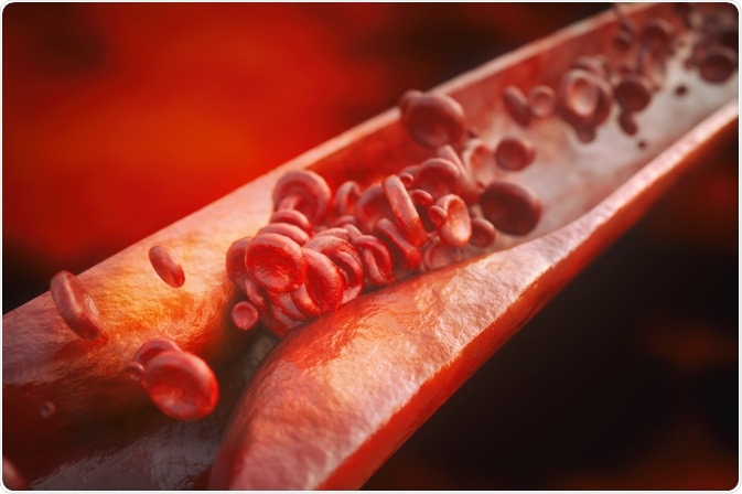
The human body is a fragile, complex system that can be afflicted by any number of diseases, pathogens, or medical conditions. The ability to image tissues and organs helps to guide improvements in drug and treatment development. Intravascular fluorescence methods are one of the latest tools which are helping to achieve this crucial medical aim.
 Image Credits: Crevis / Shutterstock.com
This article discusses intravascular fluorescence, a form of molecular imaging that uses fluorescence to examine arterial walls.
Fluorescence and fluorescence spectroscopy: an overview
Fluorescence is a phenomenon that exists in a vast range of chemical and biological systems. It occurs when atoms or molecules are subjected to light, which excites the electrons which then relax to their ground state, releasing light that can then be detected by specialized equipment. The different excitation and emission frequencies are dependent on the atom or molecule.
The phenomenon itself has been known about since the 16th Century but it was not until the publication of Sir George Gabriel Stokes’ paper “On the Change of Refrangibility of Light” that the term fluorescence entered the common scientific lexicon. Since then, fluorescence spectroscopy and microscopy methods have been developed for use in a number of studies across scientific disciplines.
It was not until the 1950s that fluorescent substances were properly reported in living systems with the discovery of green fluorescent protein (GFP) which was discovered to be a protein in 1962 by Osamu Shimomura. Since this ground-breaking research, many more fluorescent compounds (fluorophores) have been discovered and categorized. These include reactive and conjugated dyes, nucleic acid dyes, cell function dyes, and fluorescent proteins.
Technological advances in fluorescent dyes, optical instrumentation, and computer software have all contributed to the field of fluorescence imaging and our understanding of complex, dynamic biological systems and molecules in recent decades.
Fluorescence spectroscopy and associated technologies are especially suited to the study of the human body. In recent years, there have been several exciting developments that have provided medical science with the ability to improve health and life expectancy in patients.
The impact of heart disease
Many different lifestyle choices and heritable conditions can cause perturbations in the vascular system, sometimes with fatal results. Heart disease is one of the biggest killers in the world and places a huge strain on healthcare systems globally. In the US, 1 in 4 deaths annually are attributed to some form of heart disease.
Atherosclerotic lesions are an indicator of heart disease. These can lead to myocardial infarction, strokes, and critical limb ischemia (CLI.) It is of critical concern to medical scientists and healthcare professionals to provide early detection and intervention.
Atherosclerosis is an inflammatory disease caused by the upregulation of leukocyte adhesion molecules and recruitment of macrophages and T cells within the wall of blood vessels. This causes a build-up of lipids which can lead to foam cell formation and an inflammatory cascade amongst other risk factors.
Risk factors that can cause atherosclerotic lesions include high blood pressure, high cholesterol, smoking, obesity, insulin resistance, and inflammation from conditions such as lupus. The financial impact in the US alone from 2014-2015 was approximately $219 billion. Apart from the cost to the health care system alone, there is a huge cost due to lost productivity.
Lifestyle changes undoubtedly have the biggest impact in terms of preventing heart disease. However, there is a pressing need for effective and sensitive molecular imaging techniques that are non-destructive and provide data in real-time. Research in this area is key to unlocking improvements in drug delivery and effective healthcare solutions.
Intravascular fluorescence imaging: a new tool in detecting heart disease
Intravascular imaging was first introduced in the early 1990s. The technology caused a shift in our understanding and ability to identify harmful plaque build-up in vascular walls. However, there is cumulative evidence from histology-base studies and large-scale coronary atherosclerosis studies that demonstrate significant limitations in existing widely used methods.
Intravascular fluorescence is a catheter-based molecular imaging technique. The system uses near-infrared laser-induced fluorescence to image vascular walls and detect high-risk plaques. It can be used to detect either arterial wall autofluorescence or fluorescent molecules introduced intravenously.
Imaging catheters employed in this technique contain an optical fiber that delivers and collects light. These catheters are introduced through semi-invasive interventions into the target blood vessel (for example, by percutaneous coronary intervention). The fluorescence generated by the technique is then detected and analyzed by specialist imaging and data processing equipment.
Like many scientific techniques, results can be improved by using complementary techniques to provide functional information in a morphological context. These include structural imaging modalities such as intracoronary optical coherence tomography (OCT).
Studies employing intravascular fluorescence
There have been several studies carried out which have investigated the clinical applications of intravascular fluorescence. A 2016 clinical study by Verjans et al. used intravascular laser-induced fluorescence in combination with optical OCT using intravenously introduced indocyanine green to detect high-risk carotid plaques which carry a risk factor for strokes.
In 2018, Min Woo Lin and colleagues in the Republic of Korea used fluorescence lifetime imaging in conjunction with OCT to create a hybrid intravascular imaging device to visualize the structure and biochemical composition of arteriosclerotic plaques in a rabbit artery. The team studied autofluorescence of the artery: no exogenous contrast agent was used in this study.
Another interesting study which was published in 2017 by Hara T et al. in the European Heart Journal used intravascular fluorescence (using intravenously introduced molecular imaging agent FTP11-CyAm7) to detect unhealed intravascular stent in vivo. Potentially, this technique could reduce the risk of stent thrombosis and improve surgical success and recovery rates for patients.
In conclusion
Intravascular fluorescence is still a new field in clinical science and heart disease research. However, it is showing some exciting potential to become a cutting-edge part of the medical scientist’s toolkit. With an increasing number of clinical studies exploring the technology and its applications, it is likely to contribute to a new frontier in combatting one of the most persistent public health concerns in the 21st Century.
Sources
Bourantas, C.V. et al. (2017) Hybrid intravascular imaging: recent advances, technical considerations, and current applications in the study of plaque physiology Eur Heart J. 38(6) pp. 400-412 (Accessed 13th May 2020) https://www.ncbi.nlm.nih.gov/pmc/articles/PMC5837589/
Calfon, M.A. et al. (2010) Intravascular near-red fluorescence molecular imaging of atherosclerosis: toward coronary arterial visualization of biologically high-risk plaques J Biomed Opt. 15(1): 01107 (Accessed 13th May 2020) https://www.ncbi.nlm.nih.gov/pmc/articles/PMC3188610/
Min Woo Lee, et al. (2018) Comprehensive intravascular imaging of atherosclerotic plaque in vitro using optical coherence tomography and fluorescence lifetime imaging Nature Scientific Reports 8 Article No. 14561 (Accessed 13th May 2020) https://www.nature.com/articles/s41598-018-32951-9/
Tetsuya Hara et al. (2015) Intravascular fibrin molecular imaging improves the detection of unhealed stents assessed by optical coherence tomography in vivo. European Heart Journal 38(6) pp. 447-455 (Accessed 13th May 2020) https://academic.oup.com/eurheartj/article/38/6/447/2970280
Further Reading
Last Updated: Jun 19, 2020