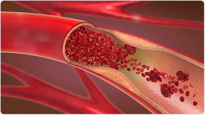
PET/angio-CT is one of the established ways to evaluate the vascular system. The method is also known as computed tomography angiography. It has been shown to be useful in tracking the progression and treatment efforts of various vascular diseases, with especial focus on inflammation and cardiovascular diseases.
 Image Credits: Christoph Burgstedt / Shutterstock.com
How does PET/angio-CT work?
PET/angio-CT is done by computer tomography and positron emission tomography, where the body is visualized. Radiotracer is injected prior to use, which then undergoes beta decay to release a positron that goes on to interact with an electron to produce two photons, detected by the scanners. The distribution of radiotracer is then evaluated.
PET/angio-CT specifically looks at the heart and blood vessels. In addition to standard PET/angio-CT, there is dynamic PET/angio-CT, which tracks movement.
The resolution of commercially available PET/CT scanners lies at around 4 mm, but those with 2 mm resolution are also available. Visualization is done by reconstruction of the collected raw data, which in dynamic PET/angio-CT is taken each 3 seconds or longer.
Using the image acquisition, image construction can be done to show 3D reconstruction of vessels. PET/angio-CT is a step up from the previously preferred method, called isotope angiography. Angio-CT has higher resolution and is more accessible, which largely accounts for its success. Other main benefits of PET/angio-CT include lower radiation exposure than some other methods, fewer artifacts, and better potential for diagnostic use.
The diagnostic performance, in particular, is an important benefit of PER/angio-CT compared to earlier methods. Compared with only PET or angio-CT alone, their use in combination has higher specificity, positive predictive value, negative predictive value, and accuracy by around 1-19% depending on performance focus.
Applications of PET/angio-CT
The most common application of PET/angio-CT is for visualizing vessels. For example, the method can be used to check whether there are any blockages in arterial vessels such as the aorta and the supra-aortic arteries going up towards the base of the skull, which provide blood supply to the brain. The detection and evaluation of coronary artery disease is a particular target of PET/angio-CT, where it is proving to be more effective than traditional methods and does not require a high radiation dose.
In addition to visualizing vessels to check for blockages, PET/angio-CT can be used to quantify blood flow. Different tracers can be used, but include 13N-ammonia, 15O-water, and 82Rb. Additionally, PET cannot alone determine the reason for changes in blood flow, which can be due, for example, to epicardial coronary artery stenosis or microvascular dysfunction, but PET/angio-CT can potentially be used to better understand underlying causes.
PET/angio-CT’s ability to visualize vascular tissue can be extended to other structures, too. Cancer has been a popular target, with tumors of melanoma, renal cell, colon, and hepatocellular carcinomas targeted. One of the most common applications is for the staging of prostate carcinoma.
PET/angio-CT is also a commonly used method to investigate cardiovascular health. For example, a PET/angio-CT image can show if atrial walls are thickening or if there is hypermetabolism of heart tissue. Thanks to its ability to pinpoint causes of stress and inflammation, the method can be used to check the correct functioning of intracardiac devices, such as automatic defibrillators, and rule out device malfunction as a cause of disease.
Limitations of PET/angio-CT
While it is a powerful visualization tool that can be carried out in many medical facilities many shortcomings of PET/angio-CT still exist. For example, PET/CT is susceptible to high costs, thereby limiting application depending on healthcare systems in place.
The main arteries, such as the femoral arteries and iliac arteries, are easily visualized. More obscured ones, such as the celiac trunk, can occasionally be visualized, whereas vessels in body parts in motion, such as the upper abdomen, can rarely be visualized and are susceptible to giving false-positive results.
The main reason for limitations in the quality of PET/angio-CT images is the long acquisition time. However, it should be noted that discrepancies in visualization can possibly be fixed with improved resolution.
Sources
Drescher, R. and Freesmeyer, M. (2013). PET angiography: application of early dynamic PET/CT to the evaluation of arteries. American Journal of Roentgenology. https://doi.org/10.2214/AJR.12.10438
Rodriguez-Garcia, J. et al. (2018). Role of PET/Angio-CT in the evaluation of intracardiac devices. https://doi.org/10.1016/j.rec.2017.05.006
Di Carli, M.F. and Murthy, V.L. (2011). Cardiac PET/CT for the evaluation of known or suspected coronary artery disease. Radio Graphs. https://doi.org/10.1148/rg.315115056
Further Reading
Last Updated: Jun 17, 2020