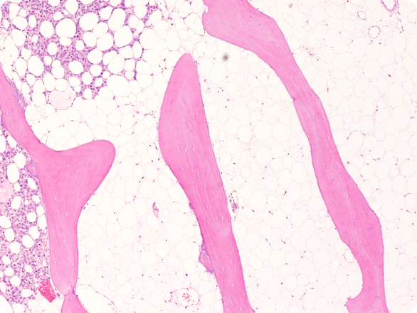
The significance of histological examination in the classification and diagnosis of clinical conditions is reliant on the expertise of the histology laboratory in managing the wide spectrum of specimen types submitted for analysis.

From receipt of the tissue sample to presentation of a slide for microscopic examination, histologists must consider the composition of the specimen to effectively determine how it should be handled.
The majority of samples follow a routine cycle of dehydration and paraffin embedding in preparation for sectioning on the microtome.
However, because of its high calcium content, bone is a particularly difficult tissue to section, with the density of a sample also being an important factor to consider in contributing to the risk of shattering. Cutting un-decalcified bone sections requires resin embedding, specialised microtomy and modified staining techniques.
In addition, an iliac bone biopsy is both procedurally very difficult to perform and uncomfortable for the patient, as it requires a 4mm core for un-decalcified processing and the sample taken must include both bony plates of the iliac crest.
As a consequence, most laboratories decalcify specimens, enabling them to be embedded in paraffin and processed as standard. Nitric acid and formic acid are used as decalcification agents, with formic acid being more suitable for laboratories performing downstream molecular analysis because, unlike nitric acid, it does not destroy DNA.
Decalcification varies in time depending on the size and type of a bone specimen. Overnight decalcification, on the other hand, is considered sufficient for a 2mm bone core biopsy and allows for subsequent IHC analysis to be performed on the sections generated.
The quality of a section can also have a significant impact on the accuracy and reliability of diagnosis, and it is dependent on selection of the most appropriate microtome.
Nigel Harness, a UK Histology Technician with 26 years of experience in sectioning bone, advocates manual sectioning for paraffin embedded specimens because of the need to use varying levels of force to cut different parts of the sample. “Manually cutting bone samples is preferable to mechanised cutting as the technician has the ability to ease off when they hit a hard area and it allows the operator to ‘feel’ how the section is being cut.”
Joint revision tissues, on the other hand, sometimes require larger and thicker sections to be cut, due to the inclusion of metal and plastics in the sample. As a result, these samples sometimes require embedding in resin with the use of a fully mechanised microtome which delivers both the controlled slow speed and constant even force needed to generate good quality sections.
Offering options for both manual and mechanised sectioning, the Thermo Scientific HM355S and Finesse ME+ microtomes provide the versatility needed by laboratories sectioning bone. Both instruments deliver a wide sectioning range, memory positioning and a choice of mechanised cutting modes, enabling laboratories to effortlessly transition between samples. Nigel stated “I’ve used a variety of different microtomes over the years but have found that the Finesse ME+ offers a good balance for our needs. It’s equally at home cutting bone as it is soft tissue and is particularly effective when it comes to ribboning.”
With its ultra-light touch flywheel, the Finesse ME+ microtome requires minimal force to operate, reducing the risk of developing RSI from prolonged use whilst, at the same time, supporting a technician’s preference to “feel” the consistency of a tissue block when sectioning.
For more information on sectioning of bone or for guidance on selecting the most appropriate microtome for your application, please contact Thermo Fisher Scientific at [email protected].