Researchers have described a new effect of stroke whereby damage to the sensory system in the affected limb means the brain develops an inaccurate map of the hand.
Publishing their case study in BMJ Case Reports, researchers examined a stroke patient’s hand using new and finer-scale methods of non-painful touch. The patient could not see the hand or the stimulus, and had to rely on information collected by tactile receptors in the hand alone to work out the exact point on the hand being touched.
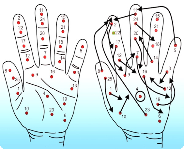
Of the 25 locations tested, the patient correctly identified the location of the touch in one instance only. Instead, when a finger was touched the patient sometimes felt the touch in the palm, adjacent fingers, or a different spot on the same finger.
“Stroke has destroyed the orderly contacts between nerve cells and resulted in the brain forming a scrambled representation of the hand,” says Dr Ingvars Birznieks, lead author of the study and a neurophsyiologist at Neuroscience Research Australia (NeuRA) and the University of Western Sydney.
“During rehabilitation the patient regained enough movement to be able to hold a shopping bag, but if she doesn’t keep her eyes on the hand while performing an action it often spontaneously releases the object. This means that the underlying problem is probably related to the body’s sensory system and not muscle force,” says Dr Birznieks.
Rehabilitation strategies to regain hand and arm function after stroke largely focus on regaining movement, however, the ability to simply move a limb is not enough as proper use of our hands relies on sensory information about the objects we touch being sent to the brain.
“This distorted view of the hand would remain undetected during routine examinations done on stroke patients, and our patient wasn’t even aware that the feelings in her hand were ‘out of place’ until our experiment,” Dr Birznieks says.
“We plan on testing more stroke survivors to find out how common this disorder is. No one currently does such tests, but they need to be done because a more accurate understanding of the underlying causes of disability following a stroke will lead to improved rehabilitation programs,” Dr Birznieks concluded.

Figure: The hand on the left indicates the 25 test sites used. The hand on the right shows the brain’s inaccurate map of the hand following stroke. Arrows originate from the centre of each test site and terminate where the stimulus was felt. Note: Site 4 was the only site where the stimulus was felt exactly where it was applied; site 22 was not felt at all; and sites 6, 15, 18 and 21 were felt but could not be localised with any confidence.