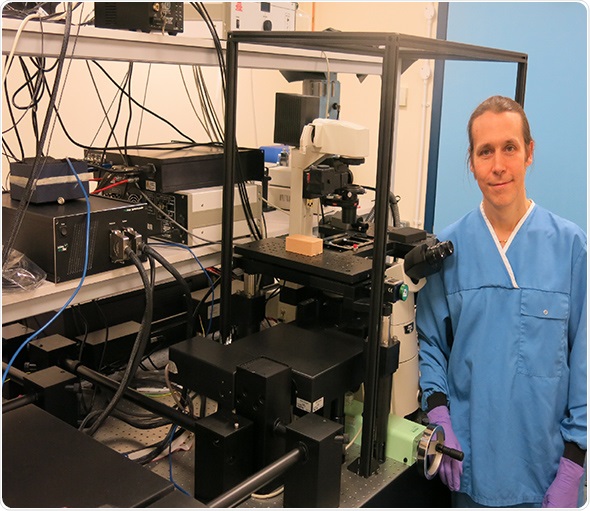
LaVison BioTec, developers of advanced microscopy solutions for the life sciences, report on the research of Professor Kurt I Anderson and his groups' studies of cell migration in the context of cancer metastasis at the Beatson Institute for Cancer Research.
Professor Kurt I Anderson is the Group Leader of the Tumour Cell Migration research program at the Beatson Institute, part of Cancer Research UK. His work focuses on developing fluorescence microscopy approaches to study the cellular and molecular dynamics of metastasis in vitro and in vivo. Metastasis is linked to mortality in most types of cancer, and a matter of intense investigation around the world. Metastatic invasion is challenging to study because it occurs randomly over large scales of time and space, and sensitively depends on features of the local tumour microenvironment.
 Speaking about his work, Professor Anderson says:
My research group studies cell migration in the context of cancer metastasis. Our research goal is to understand the influence of the tumour microenvironment on disease progression and response to therapy, especially anti-metastatic treatment
Continuing about his choice of instrument, Professor Anderson says the LaVision BioTec “TriM Scope allows us to investigate cellular and molecular interactions in 3-dimensional environments including, for example, organotypic cultures and organoids; also ex vivo and in vivo tissues. The only way for us to answer questions about the tumour microenvironment, and the effects of drugs in different regions of the tumour, is to image real mouse tumours. The only approach which allows us to do this with sub-cellular resolution of multi-photon microscopy. The TriM Scope is particularly important because it is optimised for deep tissue imaging and for fluorescence lifetime imaging (FLIM), here used to image FRET reporters which tell us about the activation state of pathways critical to the regulation of cell migration. This approach lets us map the spatial and temporal response of key signalling pathways to therapeutic intervention.”
Prior to using the TriM Scope, his group used various multiphoton & confocal systems as well as the frequency domain FLIM systems from Lambert Instruments. The TriM Scope has brought a number of benefits, as outlined by Professor Anderson.
I think the external detectors are more sensitive due to their position in direct proximity to the back focal aperture of the objective. This makes optimised custom configurations more readily available to us. Also, LaVision's experience working with OPO lasers was one of the reasons we bought their system compared to the competition. No one else has adaptive optics integrated into the scan head. We were also attracted by the cloud scanner as it offers the possibility to increase signal at intermediate levels of magnification
For more details about LaVision BioTec's TriM Scope 2-photon Microscope and its applications, please contact LaVision BioTec on +49 (0)5219151390, visit the web site: https://www.lavisionbiotec.com/.