For twenty years, it has been understood that dystrophin is expressed in differentiated muscle fibers where it is part of a protein complex that crosses the membrane and connects the extracellular matrix to the actin network inside the cell to provide structural integrity. In the absence of dystrophin, that complex is lost and movement tears the muscle membrane, leading to muscle fiber damage and repetitive cycles of repair.
This understanding is still correct and this is the driver of the muscle damage in Duchenne muscular dystrophy. Our discovery is really a paradigm shift into our understanding of why the disease is so progressive and that’s because we've discovered a new function for dystrophin in stem cells.

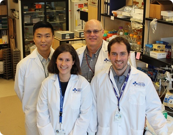
(Left to right): Will Wang, Dr. Caroline Brun, Dr. Michael Rudnicki and Dr. Nicolas Dumont
What did your recent study, published in Nature Medicine, suggest about the role of muscle stem cells?
We discovered that dystrophin plays an entirely different role in muscle stem cells. When muscle stem cells are activated, they enter the cell cycle and begin to undergo the cellular process leading to their cell division.
Stem cells can divide in two ways. They can undergo what's called a symmetric division, which results in two stem cells, or they can undergo an asymmetric cell division whereby one of the resulting daughter cells becomes a progenitor cell or what is also called a myoblast. The progenitor cell will divide several times before differentiating to repair the tissue. The other cell, of course, is the stem cell. That's why it's called an asymmetric division.
Those asymmetric cell divisions occur in a different orientation than the symmetric cell divisions. A symmetric cell division occurs parallel to the basal lamina. The basal lamina, if you can imagine, is the flooring; it’s what the cells are standing on and the two cells beside one another are both sitting on the basal lamina. The cell division plane is parallel to the basal lamina.
An asymmetric cell division occurs at a right angle to the basal lamina, so that the committed cell is on top of the stem cell. It's being pushed away from the basal lamina and towards the muscle fiber. That control of division orientation also controls whether or not the stem cell undergoes a symmetric or an asymmetric cell division.
Dystrophin is required for the cells to understand which way is up and which way is down. Dystrophin localizes to the surface of the cell that is on the basal lamina. It tells the cell where its boots are.
In the absence of dystrophin, the stem cell doesn't know which way is up and is very inefficient in performing an asymmetric cell division. Not only that, but many of the stem cells undergo centrosome amplification because they're having difficulty understanding which way is up and which way is down. This leads to what's called mitotic catastrophe and, we believe, loss of the viability of the cells. As far as we can tell, about 40% of the cells are dying via this mechanism.
Overall, the reduction of an asymmetric cell division, preferential stem cell self-renewal and loss of high numbers of stem cells leads to a reduced efficiency of stem cell function. They can still muddle through and repair the muscle, but the kinetics of repair are very slow.
Out discovery indicates that this delay and impaired regeneration process, together with repetitive injury in the muscle fibers produces the full impact of Duchenne muscular dystrophy.
%20is%20contrasted%20with%20a%20muscle%20fibre%20from%20a%20mouse%20model%20of%20Duchenne%20muscular%20dystrophy%20(bottom%20right).%20In%20normal%20mice%2c%20stem%20cells%20(pink)%20express%20dystrophin%20(green).jpg)
A normal mouse muscle fiber (top left) is contrasted with a muscle fiber from a mouse model of Duchenne muscular dystrophy (bottom right). In normal mice, stem cells (pink) express dystrophin (green) and are able to easily generate new muscle fibers, but in the disease model, there is no dystrophin and the stem cells lose their sense of direction and have trouble generating new muscle fibers. Reproduced with permission of Will Wang.
How does this finding change our understanding of Duchenne muscular dystrophy?
I think it's really a paradigm shift in our understanding. It indicates that Duchenne muscular dystrophy is a stem cell disease, as well as a muscle fiber disease.
Also, I think in a very interesting way, it explains other aspects of the disease such as the neurodevelopmental delays that occur.
Dystrophin is also involved in the heart and in the brain and so on. It's speculative right now, but conceivable, that these polarity deficits in other tissues are contributing to the full picture of the disease phenotype.
Your research was conducted in mouse cells, do you think the findings will hold in humans?
We performed very deep mechanistic studies to understand the working of dystrophin and the other polarity genes. I have a high degree of confidence that this will be found in humans because the exact same types of genes and mechanisms are present in humans.
Right now, we're working with human biopsies from donors to attempt to validate those findings. I don't see any reason why they would be different.
Is this research likely to lead to more effective treatments for Duchenne muscular dystrophy?
Knowledge is power and these are all drugable pathways. I can’t see any reason why, in the future, we could not identify novel drugs or even repurpose existing drugs to restore the function of these cells. Certainly, we've already identified a protein biologic called Wnt7a that restores the function of these cells by enforcing an alternate polarity.
Ideally, we want one small drug that does the same thing and that is the direction that our lab is moving in. Because we know about it, we can attempt to address it.
I do think it also means that for the gene replacement strategies that are being explored in trials such as exon-skipping and therapy with AAV vectors, that thought has to be given to either targeting the stem cell itself with an appropriate form of dystrophin that has its polarity function restored (these mini dystrophins that are typically being used do not have that function) or combining it with therapies that address the stem cell deficiency, together with restoring the genetic loss in myofibers.
Do you think it will be possible to ever cure Duchenne muscular dystrophy?
This is a very difficult disease and it's a hard nut to crack. I think that, further down the road, we will have therapies that will make this into a chronic disease rather than a lethal disease.
I don't know when that will occur, but I think that by combining multiple approaches to simultaneously address the genetic loss, the stem cell dysfunction, the fibrosis, and the inflammatory aspects, we will have a much better way of handling this than we do now.
What are the next steps in your research on muscle stem cells?
We are very interested in identifying drugs that ameliorate the stem cell function. We're continuing with our preclinical development of a protein biologic to do the same thing. We've made some really good progress in the lab. I'm quite excited about the future and our ability to address this stem cell deficiency.
I think this is a wonderful example of how curiosity driven research into fundamental mechanisms controlling what cells do can lead to really important insights that are going to have important clinical impact further down the road.
So often nowadays, we hear politicians and others saying that we shouldn't be funding these crazy scientists who do whatever they want; we need to have applied research. Many years ago, the director of NIH said that if we only funded applied research, we would have a very high tech iron lung for polio victims, but we would never have invented the Salk vaccine. I think this is really underscores the importance of basic research.
Where can readers find more information?
Our paper can be found in Nature Medicine
About Dr Michael Rudnicki
Dr. Michael Rudnicki is a Senior Scientist and the Director of the Regenerative Medicine Program and the Sprott Centre for Stem Cell Research at the Ottawa Hospital Research Institute. He is a Professor in the Department of Medicine at the University of Ottawa. Dr. Rudnicki is the Scientific Director of the Canadian Stem Cell Network.
Dr. Rudnicki received his Ph.D. at the University of Ottawa in 1988 with Dr. Michael McBurney where he examined the control of gene expression during embryonal carcinoma cell differentiation. Dr. Rudnicki trained at the post-doctoral level at the Massachusetts Institute of Technology in the Whitehead Institute with Dr. Rudolf Jaenisch.
His post-doctoral studies involved the genetic dissection of the function of the MyoD-family of transcription factors by gene targeting. Dr. Rudnicki was appointed Assistant Professor at McMaster University in 1992. He moved to Ottawa In 2000 to join the Ottawa Hospital Research Institute and was appointed Professor in the Department of Medicine at the University of Ottawa..
Dr. Rudnicki is an Officer of the Order of Canada, a Fellow of the Royal Society of Canada, and he holds the Canada Research Chair in Molecular Genetics. He is an Associate Editor of Cell Stem Cell and the Journal of Cell Biology, and is Co-Editor in Chief of Skeletal Muscle. He has organized international research conferences as one of the founding directors of the Society for Muscle Biology.
Dr. Rudnicki's laboratory works to understand the molecular mechanisms that regulate the determination, proliferation, and differentiation of stem cells during embryonic development and during tissue regeneration. The lab has conducted leading studies into both embryonic myogenesis and the function of muscle stem cells (satellite cells) in adult regenerative myogenesis. In particular, they have worked extensively to understand the molecular mechanisms that regulate the function of satellite cells in skeletal muscle.
Towards this end, the lab employs molecular genetic and genomic approaches to determine the function and roles played by regulatory factors. They identified Pax7 as a transcription factor required for the specification of satellite cells, and identified Wnt7a signaling as playing an important role in muscle stem cell function. Research has led to the publication of over 200 peer reviewed articles in scientific journals that include Cell, Nature, Nature Medicine, Nature Cell Biology, Cell Stem Cell and Genes & Development.