Oct 5 2016
LaVison BioTec, developers of advanced microscopy solutions for the life sciences, report on the latest work of Li Ye, a Post-Doc Research Associate in the Deisseroth Laboratory at Stanford University he applies light sheet microscopy in a program to quantitatively study brain activity in order to better understand the processing of information.
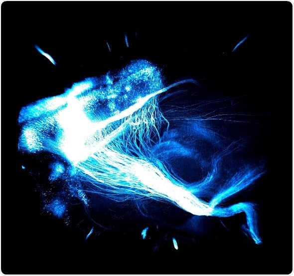
This is an image taken by the UltraMicroscope from LaVision BioTec. A 3D CLARITY volume image shows the brain-wide projection from mouse prefrontal cortex, labelled by a single stereotaxic injection of axon-filling EYFP. Image credit to Karl Deisseroth and Li Ye, Stanford University.
Dr Li Ye is post-doctoral research associate in the Deisseroth Laboratory at Stanford University. Research leader, Karl Deisseroth, is the D H Chen Professor of Bioengineering and of Psychiatry and Behavioral Sciences. They are both affiliated to the Howard Hughes Medical Institute. The Deisseroth Laboratory works on developing and applying high-resolution tools for controlling and mapping specific well-defined elements within intact and fully-assembled biological systems. The development and application of these and other tools (integrated with optical, electrophysiological, computational, molecular, and behavioral approaches) are used to study neural physiology and behavior in freely-moving mammals. The Laboratory is based on the Arastradero site where many of the group's newer technologies are being developed (this campus is part of the Stanford CNC program and the site of CLARITY development and training).
Their most recent work has developed a quantitative analysis of the wiring and molecular properties of neurons in the prefrontal cortex that are associated with distinct behavioural experiences that illuminates the logic of information processing in the brain. The work has been published online in Cell. Entitled “Wiring and Molecular Features of Prefrontal Ensembles Representing Distinct Experiences,” the paper reports on how they have developed quantitative hydrogel-based technologies to connect activity in cells reporting on behavioral experience with measures for both brain-wide wiring and molecular phenotype.
For light sheet imaging of whole mouse brains, the group uses the LaVision BioTec UltraMicroscope which provides high speed imaging and large fields of view. Talking about his use of the light sheet microscopy, Dr Ye said:
We really like the convenience and flexibility of the microscope. It has been a true workhorse for everyday sample imaging as well as for reliable data collection at the behavior-cohort scale. It works like a benchtop scanner for brains, with which you can quickly visualize the “big picture” of a whole brain in real time, yet at the same time have the option to zoom in to explore the details easily and immediately.
 Scientists show gut bacteria can reach the brain in mice and reveal a potential vagus nerve pathway