For the first time, researchers have managed to image the structure of a central component of the Ebola virus.
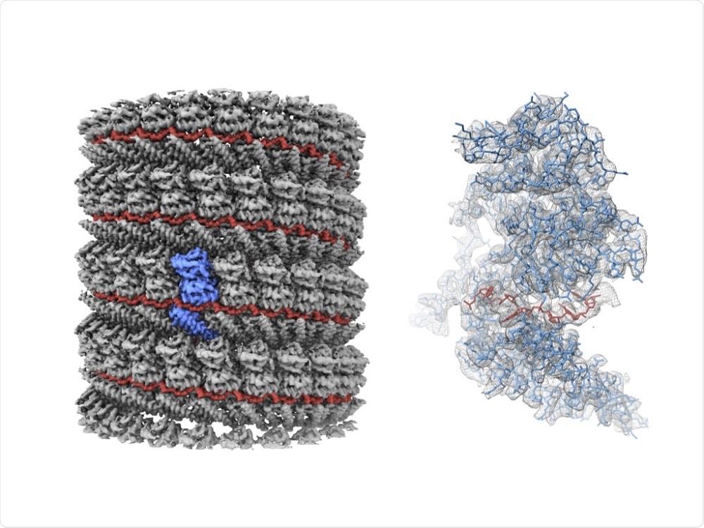
As reported in the journal Nature, Matthias Wolf and colleagues from the Okinawa Institute of Science and Technology Graduate University (OIST) have gained near-atomic resolution images of the protein complex, a development that could improve the search for treatments.
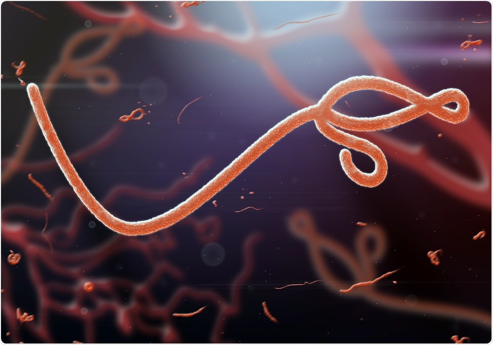
 Image Credit: jadding / Shutterstock
The team focused on the nucleocapsid; a protein complex that serves as a supporting structure for viral genetic material and enables replication.
They isolated nucleoprotein-RNA complexes that form the core of the nucleoplasmid and took the closest look yet at them using a cryo-electron microscope.
Before this study, we only knew about the smallest pieces of the NC structure. Now that we can see it as a whole, it may help find targets for antiviral drugs."
Yukihiko Sugita, Co-author
The NP-RNA complexes were engineered to lack viral RNA in non-infectious cells grown in a laboratory at OIST, meaning they did not pose any actual risk to the scientists or the public.
“Most of its components are absent. It's harmless,” says Wolf.
Previous studies have used electron tomography to analyze the nucleocapsid structure, but the cryo-electron microscopy the OIST team used achieved a much higher resolution that meant individual RNA nucleotides and amino-acid side chains could be resolved.
"Our paper shows for the first time what the NC structure looks like at near-atomic resolution" says Wolf.
However, obtaining the high-resolution images was a slow process; the protein complexes are fragileand it was difficult to get a complete sample.
It took the researchers more than 18 months to find how to reconstruct the nucleocapsid as a digital model.
Now that we have a clear picture of what this structure really looks like, it is one step closer to figuring out how the whole virus works.”
Matthias Wolf, Lead Author
The 3-D model the team has created will serve as a platform for future work, with researchers now able to perform precise and targeted studies of the whole Ebola nucleocapsid structure. The study could pave the way for new approaches to combat the virus.

Left: A 3D rendering of the cryo-EM reconstruction. The RNA and NP are colored in red and grey, respectively. A single NP molecule is highlighted in blue. Right: An atomic model of a single NP molecule with the RNA in the complex. The model is superposed with the cryo-EM map in a polygon mesh representation. (Image Credit: Yukihiko Sugita)