Researchers have successfully created miniature organoid brains in the labs that when attached to a spinal cord and muscles could help move the muscles. This could be beneficial in the research for debilitating conditions such as motor neurone disease, say the researchers. The results of the experiment were published in the latest issue of the journal Nature Neuroscience.

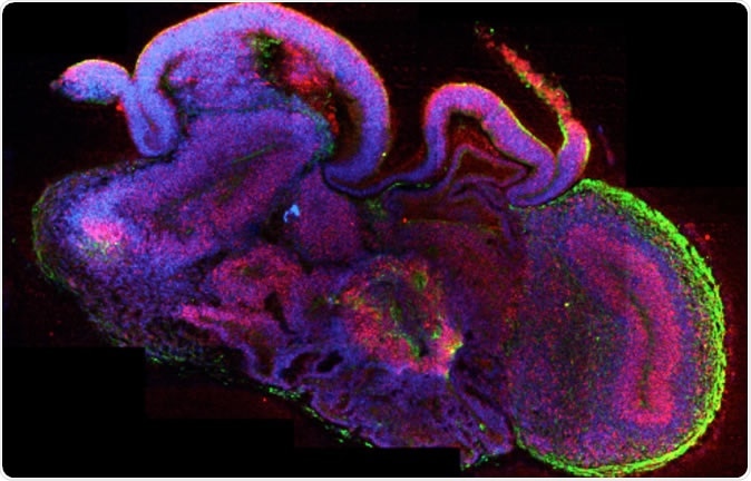
A section through a whole organoid stained for neurons in green and neural stem cells in red. All cell nuclei are stained in blue.
The small cluster of human brain cells is around the size of a lentil. When developed, it was found to develop tendril like projections and nerve connections to attach to a spinal cord and muscles from a mouse. When the brain organoid sent out signals, the muscles attached to the cord moved in response, the team noted. This is the first time that a brain organoid has been found to be attached to a central nervous system like connection and is capable of sending out distant signals, they explained. The lead author of the study, Madeline Lancaster from Medical Research Council’s Laboratory of Molecular Biology in Cambridge said, “We like to think of them as mini-brains on the move.”
For this latest experiment, the team used a new method of growth from the human stem cells. These stem cells are capable of turning into any specialized cell and it was found that these stem cells could grow into a sophisticated and complex organoid. The new organoid formed is almost as developed as a foetal brain at 12 to 16 weeks of pregnancy. It was too small to have thoughts, emotions or feelings but the signals could be generated for movement said the researchers. Lancaster said, “It’s still a good idea to have that discussion every time we take it a step further. But we agree generally that we’re still very far away from that.” She was talking about the ethical concerns of developing a living, thinking, and conscious brain in the lab.
This organoid brain contains around two million neurons, explained the team while a normal human brain contains around 80 to 90 billion neurones. This makes the organoid the size between the brain of a cockroach and a zebrafish.
This team has crossed a previously faced hurdle. Earlier development of such an organoid was complicated by the fact that nutrients would not reach the centre of the lump when it reached a particular size. This would make the inner cells die out and the experiment would be failure. This time that hurdle has been crossed. The researchers grew the brain cells and then used a thinnest of thin vibrating blades to slice the lump into thin slices – each a membrane thick. These membranes were then placed on a nutrient filled fluid and were allowed to float. The cells were thus well nourished and grew new connections and were kept alive for a year. One of the researchers, Stefano Giandomenico, developed this air-liquid interface where the slices were grown.
Once the organoid was developed, the team attached it to a 1mm-long spinal cord obtained from a mouse embryo. The brain organoid began to grow connections with the spinal cord as soon as they were placed in proximity. The cord was attached to a small bit of a back muscle from the embryo. When the organoid connected with the cord, it started sending out electrical impulses and signals that could make the muscle twitch.
Lancaster said this model could be beneficial in the research for diagnosis and treatment of several nerve diseases such as motor neurone disease, epilepsy, autism and schizophrenia. Lancaster said, “Obviously we’re not just trying to create something for the fun of it. We want to use this to model diseases and to understand how these networks are set up in the first place.”