Doctors usually diagnose patients with angina and other cardiac conditions, including coronary artery disease (CAD), through an invasive procedure called cardiac catheterization and angiography.
But, a study published in the New England Journal of Medicine, shows that instead of undergoing an invasive procedure, the patients can have a 40-minute MRI scan to detect the risk of heart attack and to determine the treatment needed.

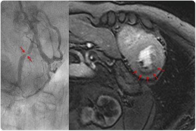
Measuring blood flow in the myocardium with magnet resonance imaging (top). The dark area in the myocardium (arrows) shows a pronounced reduction of blood flow. The cardiac catheterization of the same patient (bottom) shows a clear constriction of the artery. Image Credit: Eike Nagel, Goethe University
MRI scan vs. Angiography
The research, dubbed as the MR-INFORM clinical trial, shows that the 40-minute MRI scan to test angina can prevent patients from undergoing invasive angiography. To land to the findings, the researchers at King’s College in London analyzed more than 900 patients with angina who had two procedures, the invasive angiography or the MRI scan.
Results of the study have shown that in each group, blocked or narrow coronary vessels were dilated when indicated by the diagnostic test. Subsequently, both groups have similar outcomes, with less than 4 percent of the patients having cardiac events, including heart attack, in the following year. Also, the researchers recorded whether heart symptoms, like chest pain, persisted.
MRI: Faster and non-invasive
In the patients assigned to the MRI, group has markedly fewer procedures, and 40 percent of those patients had an invasive angiography after. Moreover, only 36 percent of the patients in the MRI group went on to have revascularization, compared to 45 percent in the other group.
This means that with the 40-minute MRI scan as the initial test, the two groups were not different with regard to symptom persistence, complications, manifestation of new symptoms, or mortality.
"This means that patients with stable chest pains who previously would have received cardiac catheterization can alternatively be examined with MRI," Professor Eike Nagel, chair in Clinical Cardiovascular Imaging at King’s College London, said in a statement.
"The results for the patients are just as good, but an examination by MRI has many advantages: the procedure takes about 40 minutes, patients merely receive a small cannula in their arm and are not subject to radiation,” he added.
The results of the study show that the MRI scan for angina is also effective in diagnosing the underlying cause of angina or chest pain. This way, the patient won’t need to undergo the invasive technique and stay in the hospital overnight.
MRI scan can also detect blockage in the coronary arteries in the heart, to determine the risk of heart attack. Hence, doctors can provide proper treatment.
The study was funded by the British National Institute of Health Research (NIHR) via the Biomedical Research Centre (BRC).
What is angina?
Angina occurs when there is decreased blood flow to the heart. Though angina is not life-threatening, it’s a warning sign that the patient is at a high risk of having a heart attack. Coronary artery disease (CAD) may cause angina. It happens when there is a blockage or narrowing of the coronary arteries, the blood vessels supplying the heart with oxygen. Atherosclerosis or the build-up of cholesterol and plaques or fatty deposits on the inner arterial wall is the major cause of CAD.
Angina also called angina pectoris, is often described as heaviness, pressure, tightness, squeezing, or pain in the chest. It is important to treat angina and to know if the coronary arteries are blocked. This way, the doctors can implement treatment, before the artery becomes totally blocked, leading to a potentially fatal condition called myocardial infarction, or heart attack.
The common signs and symptoms of angina include chest pain, pressure on the chest, feeling of fullness on the chest, pain in the arms, jaw, neck, or back, shortness of breath, fatigue, nausea, sweating, and dizziness.
Cardiac catheterization and angiography
Invasive angiography or cardiac catheterization is a procedure wherein the physician takes C-rays of the patient’s arteries. The procedure requires the patient to stay overnight. In worse cases, patients need to undergo revascularization, a procedure to improve the blood flow to the heart.
Angiography or arteriography is an imaging technique to visualize the inner part of the blood vessels in the organs of the body after the injection of a contrast medium or dye. Usually, the doctor performs the procedure to examine the veins, arteries, and heart chambers.
Sources:
- Nagel, E., Greenwood, J., McCann, G., Bettencourt, N., Shah, A., Hussain, S., Perera, D., Plein, S., Buccarelli-Ducci, C., Paul, M., Westwood, M., and Marber, M. (2019). Magnetic Resonance Perfusion or Fractional Flow Reserve in Coronary Disease. The New England Journal of Medicine. https://www.nejm.org/doi/10.1056/NEJMoa1716734
- American Heart Association (AHA). (2015). https://www.heart.org/en/health-topics/heart-attack/angina-chest-pain/angina-in-women-can-be-different-than-men