Neuroscientists have long held that the brains of children thin down over time. However, this was on the basis of older imaging techniques. Now, using cutting-edge technology in the form of quantitative magnetic resonance imaging (qMRI), a team of researchers has found that the apparent thinning is due in part to myelin formation around the cortical nerve fibers. These very interesting findings, published in the journal PNAS on September 23, 2019, will drive further research on the link between cortical function and structure.

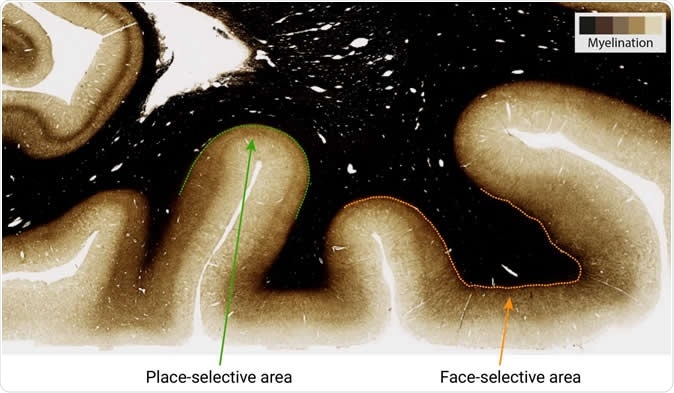
Higher myelination (darker stain) is found in the face-selective area of higher visual cortex, as compared to place selective area. Image Credit: MPI CBS
Many older studies have shown that the cerebral cortex, or gray matter of the brain, loses almost 1 mm of thickness over the period from childhood to adulthood. The gray matter is only about 3mm thick in all, so that this appears to be a considerable loss. Various explanations have emerged to account for this phenomenon. Some scientists think that the number of gray matter neurons and their projecting connections to other neurons need to be cut down for efficiency, to avoid redundancy and ensure faster connectivity. This natural process of ‘pruning’ could explain the loss as children grow and mature.
Another way to explain this is the expansion of the brain with development, as we grow. This could lead to a thinning out of the cortex as it stretches to cover a larger volume, rather like a balloon.
The new research takes the second explanation into consideration, but also draws attention to a third occurrence that has largely gone unnoticed so far, because of the limited imaging capacity of earlier measurement techniques. Using qMRI, the scientists found increased myelination occurring within the brains of younger people. Apparently this myelin caused artefacts in the assessment of cortical thickness.
What is myelin?
Myelin is the white fatty substance that encapsulates the nerve fibers, allowing nerve impulses to travel much faster by insulating most of the conductive pathway. This prevents direct continuous conduction of the impulse down the whole length of the fiber, by inducing depolarization and then repolarization at each successive point of the whole axon. Instead it forces the impulse to jump from one non-insulated gap in the myelin sheath to the next, which is artfully placed at the right distance. This is called saltatory conduction. The myelin is also a supporting structure for the delicate nerve fiber.
Previous measurements of cortical thickness have relied heavily on identifying the boundary between white and gray matter. Increased levels of myelin obscure this boundary, confusing the measurement.
The current study is based on qMRI as well as functional magnetic resonance imaging (fMRI) and diffusion MRI (dMRI). These offer independent but complementary measurements of the microstructure of the gray and white matter.
The results show that while some apparent thinning does occur, as examination of tissue samples under a microscope shows, it probably doesn’t occur to as great an extent as earlier thought. The difference is the simultaneous buildup of an insulating myelin sheath.
What did the researchers do?
In this study the scientists visualized three different areas of the ventral temporal cortex (VTC), the brain cortical area concerned with sight, in 21 children aged 5-12 years, and 30 adults aged 22-28 years. The three areas were quite close to each other, but developed in different ways. This shows that brain development cannot be interpreted in a generalized way quoting the observations made in one localized area.
They found, first, an apparent reduction in cortical thickness by 0.73 mm on average. This was not uniform with the largest reduction being 0.97 mm and the least only 0.46 mm. The difference also showed variation between left and right hemispheres though to a small extent. However, the values T1 and MD (mean diffusivity) obtained from qMRI and dMRI respectively, were consistent with tissue growth and not tissue loss. The cortical thickness was related to these MRI values in direct proportion, that is, they decreased progressively as the child grew. The change was most striking in the middle and lowest part of the gray matter, where the nerve projections are most abundant, and the adjoining white matter. This shows increased myelin content in these areas over time, as 90% of T1 variation in white matter is due to myelin growth. However, based on prior research, the researchers also point out that gray matter such as glia, synapses, and dendritic branches also contribute to tissue growth.
In the current case, the areas responsible for recognition of faces and words were myelinated. On the other hand, the area responsible for place recognition was not myelinated, but did show a stretching effect to become thinner over time. Thus the structure of the cortex in different places is linked to its function. To confirm this, the brains of adults were subjected to post-mortem examination with both ultra-high field MRI and tissue examination under a microscope.
Importance of the study
Such findings are not easy to accept as they overturn decades of accepted research findings. There is a lot of older research that indicates cortical thickness changes as one learns a new skill. Now this will be disputed, since the change could be partly due to myelination occurring at the same time. Previous research will need to undergo re-evaluation to confirm the reliability of the measurements.
However, qMRI also offers the potential to improve the detection of conditions like multiple sclerosis which are caused by the breakdown of myelin over the nerve fibers, leading to impaired nerve impulse transmission. The treatment outcomes can also be monitored with greater accuracy using this technology, which promises to be a valuable research and treatment tool in neurology.
Journal reference:
Apparent thinning of human visual cortex during childhood is associated with myelination. Vaidehi S. Natu, Jesse Gomez, Michael Barnett, Brianna Jeska, Evgeniya Kirilina, Carsten Jaeger, Zonglei Zhen, Siobhan Cox, Kevin S. Weiner, Nikolaus Weiskopf and Kalanit Grill-Spector. Proceedings of the National Academy of Sciences. 23 September 2019. https://doi.org/10.1073/pnas.1904931116. https://www.pnas.org/content/early/2019/09/19/1904931116