A new study presented at the Annual Meeting of the American Academy of Ophthalmologists (AAO) on October 14, 2019 reports an astonishing 95.% yield when artificial intelligence (AI) is used to screen a real life population for diabetic retinopathy. Not only is this amazingly sensitive, but it requires no trained ophthalmologist to perform the screening, and the results are available in just one minute flat. This AI real-time screening is ideal for primary care facilities and for the most sophisticated specialist diabetic care centers alike. The benefit of the technology called EyeArt is its potential to help doctors to quickly screen out those patients who need referral to specialized eye services to prevent permanent visual loss due to diabetes.
The need
Diabetes mellitus is a metabolic condition affecting hundreds of millions of people the world over. In just the USA, there are over 30 million diabetics. Characterized by abnormal regulation of blood glucose levels, diabetes causes not only potentially fatal alterations in the fluid balance of the body, but deprives cells of their energy supply, causes vascular injury, and increases the risk of cardiovascular disease. Its vascular complications can lead to disease of the retina, kidneys, heart and brain. A full 25% of patients with diabetes can expect to develop diabetic retinopathy, and in the USA, with the eradication of many other causes of visual impairment, this has become the No. 1 cause of blindness in working-age people.

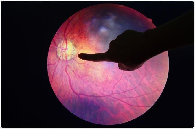
Diabetic Retinopathy. Image Credit: Anukool Manoton / Shutterstock
The process of retinal deterioration in diabetes is a prolonged one, but is hastened and aggravated by poor blood glucose control. The mechanism of damage is mediated by the increased presence of glucose in the blood. This causes direct endothelial injury, the endothelium being the smooth flat cellular lining of the interior of every blood vessel.
The weakening of the blood vessels in the eye causes small bulges to occur, between adjacent endothelial cells. This disrupts the endothelial barrier, allowing blood cells and serum to leak out into the retina outside. The intense inflammatory reaction that occurs in response to this leakage produces edema which is most marked in the fovea, the point where the retina focuses with greatest sharpness.
While early diabetic retinopathy may not cause symptoms, over time the visual impairment increases and it finally results in blindness. There are now treatments to arrest and correct the visual loss associated with this condition, provided it is caught early. For this reason, annual screening for diabetic retinopathy is an essential part of diabetes care. The present approach uses an automated approach which achieves a high degree of accuracy while screening a high volume of patients with great rapidity.
About EyeArt
An AI-based system called EyeArt was developed some years earlier, but its clinical validity remained unproven. It uses algorithms to analyze retinal images along with deep machine learning to detect and locate diabetic retinopathy lesions.
The current study aimed to assess the clinical accuracy of this screening system as compared with ETDRS, a gold-standard grading system used by experts.
EyeArt in 60 Seconds : Diabetic Retinopathy Screening Technology
Study outcomes
The study included about 900 patients in 15 different centers, who were screened using the EyeArt program on undilated eyes (without the use of dilating eye drops). These results were then sent to certified ETDRS graders for expert review. The graders found that 95.5% of eyes with diabetic retinopathy were correctly identified using EyeArt, while 86% of eyes classified as normal by the system were truly unaffected. A very small percentage of eyes could not be classified correctly without dilation. When these eyes were also included in the study, more eyes could be graded correctly. Overall, 90% or more of all eyes picked up as abnormal by EyeArt had either diabetic retinopathy or another disease of the eye.
The current investigation followed up on an earlier retrospective study using data from 100 000 successive patient visits, which found that diabetic retinopathy cases were picked up early by EyeArt, giving it a sensitivity and specificity of over 91%. The images used were those captured by real-life clinics. Validation has also been performed independently by the UK NHS in more than 20 000 patients. Images or poor quality, or those which do not show the required retinal fields, are automatically tagged by the program. This occurs within a minute of receiving such ungradable images, so that a repeat photograph can be acquired during the same patient visit, increasing convenience and patient compliance. The software is compatible with a wide range of camera models as well. Data transmission and storage follow encrypted protocols to keep all patient information private and secure, but also allows its incorporation into existing electronic healthcare record systems.
The importance
Researcher Srinivas Sadda says, “Accurate, real-time diagnosis holds great promise for the millions of patients living with diabetes. In addition to increased accessibility, a prompt diagnosis made possible with AI means identifying those at risk of blindness and getting them in front of an ophthalmologist for treatment before it is too late.” The process is as simple as taking a color image of the retina of the patient, uploading it for cloud-based analysis, and receiving the reports within a minute.