In 2019, the emergence of mysterious lung disease in the United States has sparked global health attention. Now named as the e-cigarette, or vaping, product use–associated lung injury (EVALI), the disease causes lung damage, which is a serious and sometimes fatal complication of vaping.
A team of researchers at UC San Diego Health emphasized the importance of pulmonary imaging in the diagnosis of acute lung injury linked to vaping. The special review, published in the journal Radiology, shows what is currently known about the illness and sheds light on the role of radiologists in the evaluation of suspected EVALI.
What we know
EVALI is a type of lung injury or damage linked to the use of e-cigarettes, which gained popularity about a decade ago as an alternative to cigarette smoking. In August 2019, however, cases of a mysterious lung disease emerged in the United States.
The U.S. Centers for Disease Control and Prevention (CDC) reports that as of Jan. 14, a total of 2,668 patients were hospitalized due to EVALI from all 50 states, two U.S. territories, and the District of Columbia, and there had been 60 deaths as of writing.
The CDC noted that all EVALI patients have admitted using an e-cigarette or vaping products. Further, it reports that a recent study analyzed samples from 51 EVALI cases in 16 states and found that vitamin E acetate was identified in bronchoalveolar lavage fluid samples. Vitamin E acetate has been strongly linked to the outbreak of lung injury. However, there isn’t sufficient evidence to rule out other chemicals that can cause lung illness.
Role of pulmonary imaging
Pulmonary imaging has helped a lot in detecting lung injury related to vaping. Radiologists also play a pivotal role in evaluating suspected EVALI cases. If detected early, doctors can initiate prompt treatment, reducing the risk of severe lung injury and boosting chances of survival and recovery.

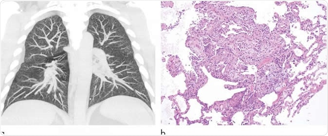
Images show electronic cigarette or vaping product use-associated lung injury in a 32-year-old man with history of vaping who presented with fevers and night sweats for 1 week. (a) Coronal maximum intensity projection image shows diffuse centrilobular nodularity. (b) Histologic sections of his transbronchial cryobiopsy showed distinctive micronodular pattern of airway-centered organizing pneumonia, corresponding to centrilobular nodularity seen at CT. Similar imaging and pathologic findings have been described in patients with smoke synthetic cannabinoids. Image Credit: Radiological Society of North America
“Rapid clinical and/or radiologic recognition of EVALI allows clinicians to treat patients expeditiously and provide supportive care. Although detailed clinical studies are lacking, some patients with EVALI rapidly improve after the administration of corticosteroids. Additionally, making the correct diagnosis may prevent unnecessary therapies and procedures, which themselves can lead to complications,” Dr. Seth Kligerman, division chief of cardiothoracic radiology at UC San Diego Health and associate professor at UC San Diego School of Medicine.

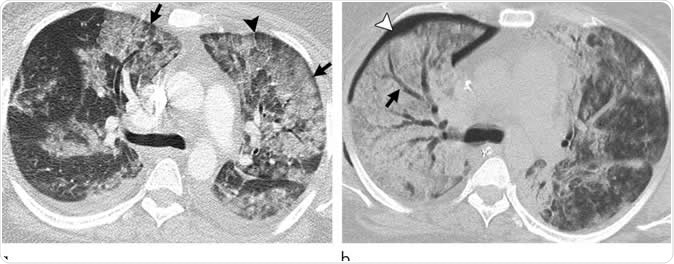
Images show electronic cigarette or vaping product use-associated lung injury with diffuse alveolar damage pattern in a 35-year-old woman who vaped tetrahydrocannabinol. Work-up for infection and rheumatologic disease was negative. (a) Axial CT scan shows ground-glass opacity, left greater than right, with areas of consolidation. Subpleural and perilobular sparing is present (arrows). Septal thickening is present (arrowhead). (b) CT scan 2 weeks later shows extensive right lung consolidation with areas of bronchial dilation (arrow) and internal development of right pneumothorax (arrowhead). Ground-glass opacity in left lung has improved with residual centrilobular nodularity. Patient died 5 days later. Image Credit: Radiological Society of North America
Though an investigation is still ongoing, the exact cause of EVALI remains unclear. Though a majority of the patients or more than 80 percent reported the use of tetrahydrocannabinol (THC) or cannabidiol CBD containing compounds. Further, most patients were adolescent and adult men.
Those with EVALI presented with symptoms such as fatigue and fever, along with respiratory symptoms and gastrointestinal symptoms, including cough, shortness of breath, the difficulty of breathing, chest pain, unexplained weight loss, vomiting, and diarrhea.
Pulmonary imaging has provided doctors with a view of the extent of lung injury. In most chest CT findings, patients showed a pattern of lung injury with sparing of the periphery of the lungs. Before diagnosing the patients with EVALI, they should have a history if vaping or e-cigarette use within 90 days and abnormal chest imaging results. Other causes of possible lung disease should be eliminated for doctors to diagnose the patient of vaping-related lung illness.

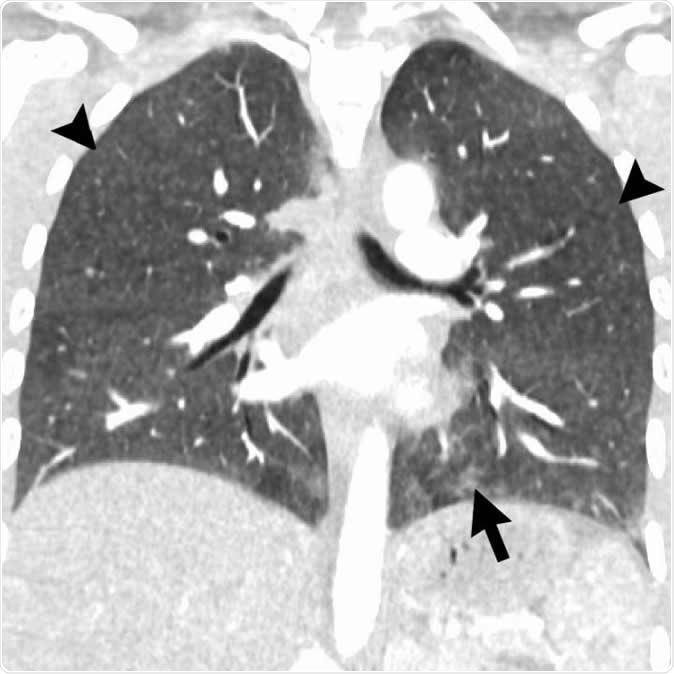
Coronal image shows hypersensitivity pneumonitis (HP) pattern in a 35-year-old man who vaped tetrahydrocannabinol products. Extensive hazy centrilobular nodularity (arrowheads) is most pronounced in midlung and upper lung zones consistent with inhalational injury. Mild ground-glass opacity is present as bases (arrow). This imaging pattern is commonly seen in HP. Patient's condition rapidly improved after steroid administration and no biopsy was obtained. Although authors have seen a few cases with HP pattern, there are no cases in literature with pathologic confirmation. Other possible etiologies for diffuse pattern of centrilobular nodules in electronic cigarette or vaping product use-associated lung injury includes airway-centered foci of organizing pneumonia. Image Credit: Radiological Society of North America
Early detection, early treatment
In some cases of EVALI, patients show mild symptoms or radiologic findings in the emergency department. The authors of the study noted that if the condition is not diagnosed in a timely manner, the patients may continue vaping, adding more injury to the lungs.
Detecting the condition early can help doctors decide on the best course of treatment for a better prognosis. Also, EVALI is just one of the string of complications linked to vaping. E-cigarette use has been tied to other conditions and long-term health risks, including THC and nicotine addiction, chronic pulmonary injury, and cardiovascular disease.
Journal reference:
Radiologic, Pathologic, Clinical, and Physiologic Findings of Electronic Cigarette or Vaping Product Use–associated Lung Injury (EVALI): Evolving Knowledge and Remaining Questions Seth Kligerman, Costa Raptis, Brandon Larsen, Travis S. Henry, Alessandra Caporale, Henry Tazelaar, Mark L. Schiebler, Felix W. Wehrli, Jeffrey S. Klein, and Jeffrey Kanne, https://pubs.rsna.org/doi/10.1148/radiol.2020192585