Breast reconstruction after removal or mastectomy of the breast for cancer is a common procedure among breast cancer survivors. In a new development, surgeons have developed a process to map out the blood vessels in the tissues of the abdomen that would be used to reconstruct the breast, prior to the mastectomy.
This mapping would help the researchers predict the chances of the graft using the tissues being successful. The team from the UT Southwestern Medical Center published the results of their study that could benefit hundreds of thousands of women undergoing breast reconstruction surgery, in the latest issue of the Journal of Reconstructive Microsurgery.

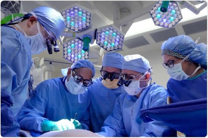
Surgeons at UT Southwestern have developed a process to determine the best approach for single breast reconstruction. Image Credit: UTSW
The surgeons explained that when abdominal tissues are used to reconstruct single breast after a mastectomy. There is a high risk of complications. These complications arise due to lack of blood flow to the whole reconstructed breast. As blood flow becomes scarce in certain regions, there is resultant fat necrosis that can lead to pain and lump formation and ultimately tissue death and failure of the reconstruction due to infections and scarring. In order to overcome these complications, the team said that if they could map out the blood flow pattern of the donor site tissue, they could predict the possible outcome of the reconstruction surgery.
The team said that before the mastectomy, the patients are routinely advised radiographic imaging studies such as computed tomographic angiography (CTA). When extended to donor sites over the abdominal tissues, these radiographic images can provide a detailed picture of the network of blood vessels, they wrote. If the surgeons are aware of the location and density of the blood vessel network in the abdominal tissue, they are also likely to assess the blood vessels they would need for the reconstruction surgery. This can help them plan the surgery in a more efficient way they wrote. The blood vessels, they wrote would be richly supplying the pedicle flap that would be used to reconstruct the single breast and this would shape the breast into its best form with least risk of complications, they wrote.
Sumeet S. Teotia, lead author of the study and associate professor of plastic surgery at UT Southwestern Medical Center and director of the breast-reconstruction program at UT Southwestern’s Harold C. Simmons Comprehensive Cancer Center, in a statement said, “Now we use the CTA results to have a conversation with the patient in clinic. Reviewing the scans helps the patient see what we may encounter in surgery and gives them confidence going into the procedure. Potentially, we can reduce and even avoid long-term complications.”
They explained that single breast reconstruction, using the tissues from the abdomen has been a challenge because the tissues often fail to recreate the normal shape and size of the breast. Those with inadequate abdominal tissues, or scars over the abdomen are also difficult candidates for such reconstructive surgeries say experts. In these cases there is not enough tissue to recreate the breast. If the other normal breast sags or is large, the reconstruction of the single breast with available abdominal tissue becomes more of a challenge. Despite these, one of the biggest problems till date wrote the researchers, is the likelihood of getting inadequate blood supply to the reconstructed breast. This often results in failure of the surgery.
For this study they looked at free flap surgical reconstructions of the breast performed on patients at UT Southwestern between 2009 and 2017. They could find 512 such patients and 150 of these patients were included in this study and followed up for results of the surgery. These women were of an average age of 57 years and had an average Body Mass Index (BMI) of 27. They found that 75 of these women had undergone single breast reconstruction while 75 underwent “conjoined, stacked double-pedicle flap reconstruction”. They also noted that 81 percent had to undergo delayed reconstruction of the breast.
The results showed of all of these cases that the women who had undergone conjoined stacked double-pedicle flap reconstruction needed lesser number of blood vessels for their flap and also had a lower risk of fat necrosis (at 2.7 percent cases). Among women who underwent a single flap reconstruction, the risk of fat necrosis was as high as 14.4 percent, the team found.
Teotia said, “The CTA scan allows us to visuospatially create a model of the breast in advance so we can be faster in the operating room. The surgeons still visually confirm the scan results during surgery, assuring a high degree of reliability.” He added, “Our algorithm allows us to be anatomically strategic in choosing the best blood supply for that particular tissue; it is important to have optimal blood supply for the best results, which ultimately translates into an improved outcome and breast shape.”
Journal reference:
Min-Jeong Cho, Sumeet S. Teotia, Nicholas T. Haddo, Classification and Management of Donor-Site Wound Complications in the Profunda Artery Perforator Flap for Breast Reconstruction, J reconstr Microsurg 2020; 36(02): 110-115, DOI: 10.1055/s-0039-1697903, https://www.thieme-connect.de/products/ejournals/abstract/10.1055/s-0039-1697903