Coronaviruses have 'spilled over' to human populations throughout history, causing respiratory illnesses, often associated with pneumonia. The severe acute respiratory syndrome (SARS), which emerged in 2002 in China, and the Middle East Respiratory Syndrome (MERS), which was first seen in Saudi Arabia in 2012, are similar to the current global pandemic spreading worldwide, the coronavirus disease (COVID-19).
For most patients who succumbed to COVID-19, the ultimate cause of death was pneumonia, a condition that causes inflammation and fluid accumulation in the lungs, making it hard to breathe. Some patients develop acute respiratory distress syndrome (ARDS), a potentially fatal condition where the lungs cannot provide enough oxygen to the vital organs of the body.
Severe pneumonia requires extended hospital stays, and, in some cases, patients are placed in intensive care units (ICU) with mechanical ventilation to help them breathe. As the COVID-19 ravages across 184 countries and territories, there has been a shortage of mechanical ventilation, and many healthcare systems are overwhelmed.
It is, therefore, crucial to determine which patients need more supportive care in hospital and those who can recover at home. To help detect which patients require more medical care, University of California San Diego Health radiologists and doctors are now using artificial intelligence (Ai) to analyze lung imaging as part of a clinical research study.
The researchers emphasized that they are not using lung imaging analysis to diagnose COVID-19, but providing a triaging method for healthcare providers to spot presumptive cases that need immediate medical care. The only way to diagnose the coronavirus disease is when a patient tested positive in the accredited tests that detect the SARS-CoV-2 or the severe acute respiratory syndrome coronavirus 2, the causative agent of COVID-19.

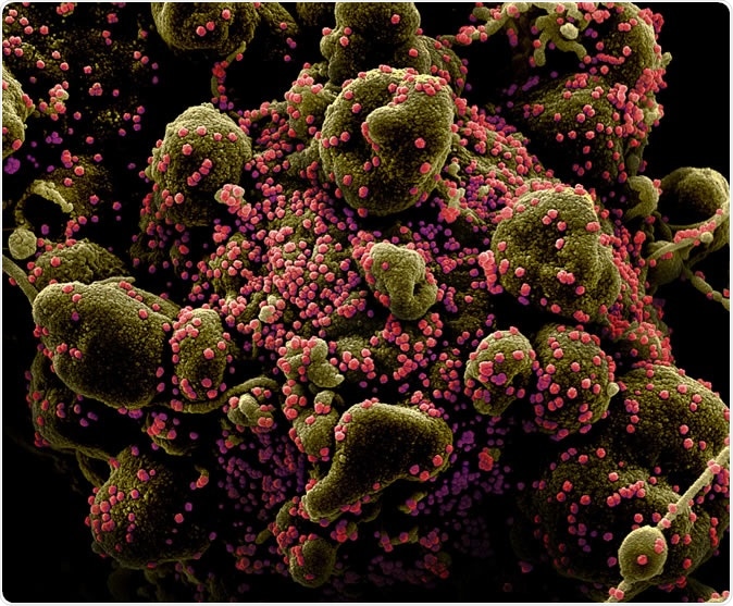
Novel Coronavirus SARS-CoV-2 Colorized scanning electron micrograph of an apoptotic cell (greenish brown) heavily infected with SARS-COV-2 virus particles (pink), isolated from a patient sample. Image captured and color-enhanced at the NIAID Integrated Research Facility (IRF) in Fort Detrick, Maryland. Credit: NIAID
Promising AI method
The novel artificial intelligence method has provided the physicians in UC San Diego Health with unique information from a bank of more than 2,000 images. For instance, a patient admitted in the Emergency Department had no symptoms of COVID-19 but had a chest X-ray for other purposes. When the image had been analyzed using the AI method, it appeared that the patient had early pneumonia. The patient was then tested for COVID-19, and it returned positive.
The AI method does not only detect those who have severe pneumonia or those who may be carriers of SARS-CoV-2 but also those who are asymptomatic but are developing early pneumonia. Treating pneumonia early is crucial to saving lives and reducing the burden on intensive care units.

Chest X-rays from a patient with COVID-19 pneumonia, original x-ray (left) and AI-for-pneumonia result (right). Patient has a pacemaker device and an enlarged heart, which indicates that the AI algorithm is powerful enough to work even when the patient has underlying health issues.
“We would not have had reason to treat that patient as a suspected COVID-19 case or test for it if it weren’t for the AI. While still investigational, the system is already affecting clinical management of patients.” Dr. Christopher Longhurst, chief information officer, and associate chief medical officer for UC San Diego Health, said.
The AI method kickstarted months ago when Dr. Albert Hsiao, an associate professor of radiology at UC San Diego School of Medicine, and his team developed a new machine-learning algorithm to permit radiologists to utilize AI to improve their ability to detect pneumonia on chest X-rays.

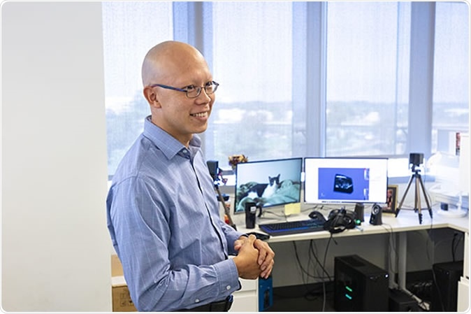
Albert Hsiao, MD, PhD, associate professor of radiology at UC San Diego School of Medicine and radiologist at UC San Diego Health, and team developed a machine learning algorithm that allows radiologists to use AI to enhance their own abilities to spot pneumonia on chest X-rays.
How does it work?
The new algorithm overlays X-rays with color-coded maps to detect pneumonia. It has been trained with about 22,000 notations by human radiologists. To test their algorithm, the team used the artificial intelligence approach to ten chest X-rays from five patients in China and the United States with COVID-19. The ten images were published in medical journals.
Even if the images were taken at different hospitals, the algorithm was able to detect localized areas of pneumonia consistently. Published in the Journal of Thoracic Imaging, the results show that the new method can detect pneumonia in patients rapidly, providing prompt treatment.
“Pneumonia can be subtle, especially if it’s not your average bacterial pneumonia, and if we could identify those patients early before you can even detect it with a stethoscope, we might be better positioned to treat those at highest risk for severe disease and death,” Hsiao said.
The researchers said that chest X-rays are more affordable, with the machines easier to clean and move around. Further, the results return faster than any other diagnostic imaging tests. Further, it is safer and emits less radiation for patients, making it a rapid test that can help fast track detection of pneumonia for proper treatment.
Source:
Journal reference: