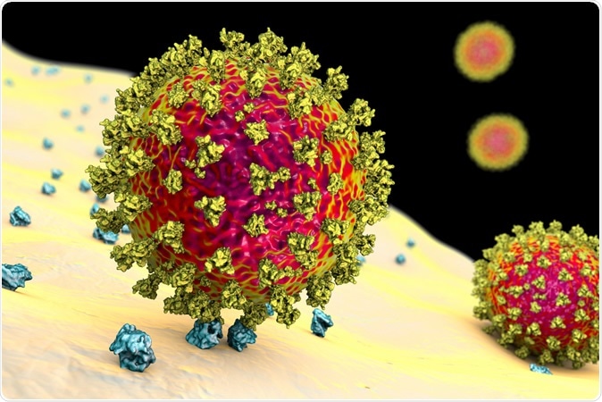
The virus has caused over 400,000 cases and 280,000 deaths in just over four months, causing immense stress in many countries and forcing nation-wide lockdowns in others. SARS-CoV-2 enters the host cell through the angiotensin-converting enzyme 2 (ACE2), which are, therefore, the viral receptors. Another novel receptor called CD147 or Basigin, has also been reported.

SARS-CoV-2 viruses binding to ACE-2 receptors on a human cell, the initial stage of COVID-19 infection. Image Caredit: Kateryna Kon / Shutterstock

 This news article was a review of a preliminary scientific report that had not undergone peer-review at the time of publication. Since its initial publication, the scientific report has now been peer reviewed and accepted for publication in a Scientific Journal. Links to the preliminary and peer-reviewed reports are available in the Sources section at the bottom of this article. View Sources
The virus attaches to the receptor through the spike or S protein, which exists as a trimer, with two subunits that are cleaved by the host cell transmembrane protease serine 2 (TMPRSS2) on the host cell upon viral attachment.
This cleavage leads to the dissociation of the S1 subunit and the fusion of the virus with the membrane, leading to virus entry and cell infection. Two other enzymes called cathepsin B, and L are able to cleave the S protein as well, though less efficiently.
Tracing vulnerability to SARS-CoV-2 at the molecular level
The current study uses the level of expression of these molecules across multiple tissues to identify which cell types could be susceptible to infection by SARS-CoV-2. Several earlier studies looking at ACE2 and TMPRSS2 expression in a number of tissues have found the organs at risk of serious damage and the target cell types. These include vital organs like the heart, kidneys, lungs and small intestine, and the type II alveolar (lung) cells, myocardial (heart muscle) cells, and epithelial cells in the ileum of the small intestine.
If Basigin is also a viral receptor, the virus may also infect T cells which have this molecule on their surface. At the same time, studies show that besides the most studied respiratory droplet route, the ocular route is also a potential means of transmission for SARS-CoV-2. This finding was extended to the current virus as well, with its ability to infect conjunctival cells in vivo and replicate.
These cells also carry high levels of ACE2 and TMPRSS2, as well as Basigin. Other researchers have reported the presence of the virus in ocular fluids with resulting conjunctival inflammation. If so, it could lead to respiratory disease via the nasolacrimal duct.
Creating a cell atlas for the eye
These findings incited interest in finding the mechanism of ocular infection by SARS-CoV-2. In order to do this, the researchers used single-cell analysis to prepare a high-resolution profile of the RNA transcriptional products of almost 33,500 single cells from human ocular tissue, creating an atlas of the eye.
In the next step, they analyzed gene expression patterns across tissues and cell types in the human eye. They found that six percent of corneal cells show a high percentage of expression. These are mostly epithelial cells, found in both cornea and conjunctiva.
Both ACE2 and TMPRSS2 are co-expressed in conjunctival cells and the former in some epithelial cells, which also express the cathepsins B and L. This may indicate the susceptibility of these cells to SARS-CoV-2 infection.
They also looked for genes encoding proteins required for the entry of other types of viruses and found that though MERS virus entry genes were not specifically found, those linked to Influenza virus entry were present, which confirms the known pattern of MERS not causing ocular complications while influenza does.
They then searched for genes that are often found along with ACE2/TMPRSS2. The list was analyzed in terms of gene function, when most of them were found to be related to cell membrane processes, including cadherin binding, cell-cell junctions, and endosomal complex. Many of these were, therefore, potentially helpful to the viral replication cycle.
Potential mechanism of SARS-CoV-2 infection through the eye
Following one such highly correlated gene, called SOX13, a transcription factor that is active in cells bearing ACE2, as well as its upstream regulator, STAT1, they found that cells that express both of these at high levels also express ACE2. The presence of SOX13 with its associated genes that modulate immune signaling after viral entry or infection indicates its potential role in ocular infection with this virus.
SOX13 could thus be targeted to reduce SARS-CoV-2 expression.
Interestingly, the researchers found that IL6, a proinflammatory cytokine that is high in COVID-19 patients with severe infection, is stimulated by endothelial cells that have receptors for interferons released by infected corneal and conjunctival cells. IL-6 activates other cellular interferon pathways associated with inflammation, while ACE2 suppresses them, providing a tight balance between them.
The findings thus show a potential pathway for the entry of and infection with SARS-CoV-2 in human ocular tissue that could spark research into the importance of this route of spread. It could also serve as a base for developing new treatments for this novel virus.
Video of Coronavirus SARS-CoV-2 structure
Coronavirus SARS-CoV-2 structure

 This news article was a review of a preliminary scientific report that had not undergone peer-review at the time of publication. Since its initial publication, the scientific report has now been peer reviewed and accepted for publication in a Scientific Journal. Links to the preliminary and peer-reviewed reports are available in the Sources section at the bottom of this article. View Sources
Journal references:
- Preliminary scientific report.
Hamashima, K., et al. (2020). Potential modes of COVID-19 transmission from human eye revealed by single-cell atlas. bioRxiv preprint. doi: https://www.biorxiv.org/content/10.1101/2020.05.09.085613v2
- Peer reviewed and published scientific report.
Gautam, Pradeep, Kiyofumi Hamashima, Ying Chen, Yingying Zeng, Bar Makovoz, Bhav Harshad Parikh, Hsin Yee Lee, et al. 2021. “Multi-Species Single-Cell Transcriptomic Analysis of Ocular Compartment Regulons.” Nature Communications 12 (1): 5675. https://doi.org/10.1038/s41467-021-25968-8. https://www.nature.com/articles/s41467-021-25968-8.