The COVID-19 pandemic caused by the severe acute respiratory syndrome coronavirus 2 (SARS-CoV-2) has stimulated intensive research into various aspects of viral infection, adaptation, and pathogenesis. One recent study by University of Pittsburgh researchers and published on the preprint server bioRxiv* in June 2020 reports the results of growing and characterizing the virus, as well as the development of neutralizing antibodies in the serum of acutely infected hospitalized patients with COVID-19.
The viral genome encodes both structural and non-structural proteins. Among them are the nucleocapsid (N), the spike (S), membrane (M), envelope (E), and various accessory proteins. These have various functions, with the viral capsid being composed primarily of N protein, which also contributes to viral replication, transcription, and viral assembly. It also produces adaptive changes in the host cell environment.
The S protein is a fusion protein that mediates viral attachment to the human ACE2 receptor on the cell membrane, which triggers viral-cell membrane fusion to facilitate viral entry into the cytoplasm. Two cleavage steps are essential for this to happen.
The cleavage enzymes are usually supplied by the host cell, and gives rise to the S1 protein that is responsible for viral attachment, and give rise to the S2 protein, which mediates fusion after further processing. Some coronaviruses are primed by cleavage during viral synthesis in the infected cell. However, the S protein of SARS-CoV-2 is cleaved only after the first generation of replicated viruses from the infected cell attach to and enter the next cell.

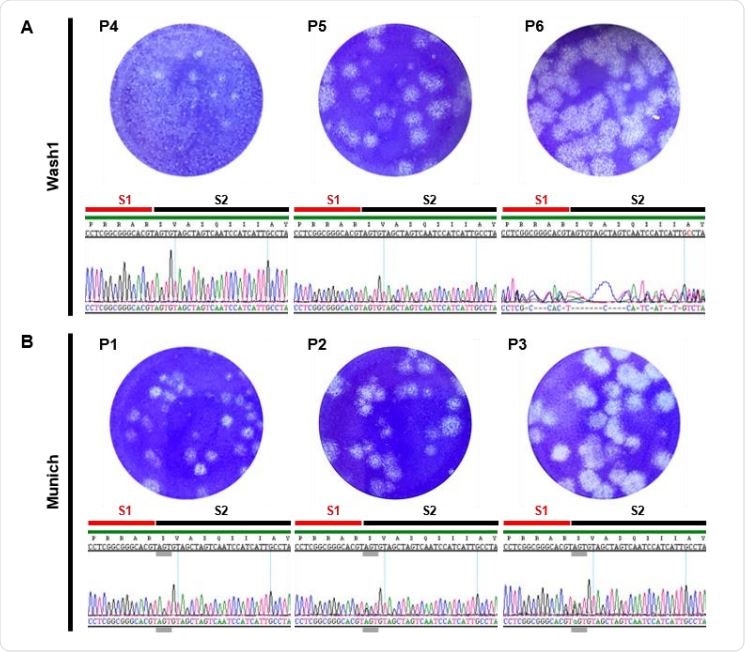
Virus growth and characterization. (A and B) Representative images showing plaques generated by infection of Vero-E6 cells with different passages of the Wash1 (A) and Munich (B) virus stocks. Chromatograms under each plaque image show the nucleotide sequence coding for the furin cleavage signal (RRAR) region of the S protein for each virus passage. Red and black lines indicate the putative S1 and S2 subunits, respectively, generated after cleavage. Grey lines indicate the nucleotide triplet coding for S 686.
Variety of Proteases
There are a variety of proteases that cleave the S protein in different contexts, such as the proprotein convertases, cell surface proteases or lysosomal proteases, and furins, depending on the time of spike cleavage. Furins act during virus packaging, elastases after virus release, TMPRSS2 enzyme after virus attachment, and cathepsin L after viral endocytosis to enter the host cell.
The SARS-CoV-2 S protein has a novel insertion at the S1/S2 interface, that is not seen in the bat reservoir virus, and this is supposed to signal a furin cleavage signal. Researchers found that furin cleavage is necessary to facilitate the entry of the pseudovirus expressing the SARS-CoV-2 S protein into other cell lines that express the human ACE2 receptor, and for the infection of human lung cells by the SARS-CoV-2 virus.
The S protein is a prime target for developing vaccines and for the natural neutralizing antibody response. Most CD4 and CD8 T cells are directed at the S, M, and N proteins.
Virological and Immunological Characterization
The current study aimed at analyzing the viral isolates from two patients, in terms of viral characteristics and immunological response, as well as to observe the changes in the furin cleavage site genotype after passage through Vero cell culture. The researchers also looked for changes in the neutralizing antibody response to changes in genetic sequence at this region.
Finally, they looked at the neutralization antibody titer in serial samples in acute COVID-19 infection. The study thus addresses significant unknowns in the standardization of virus strains, the interaction between cells in vitro, the pathogenesis of the disease, and the best strains for vaccine and drug testing.
Mutations and Consequences
The two virus strains selected for passaging represented European and American isolates. The researchers found, firstly, that the American Wash1: P6 (passage 6) isolate had a deletion mutation in the sequence encoding the furin cleavage signal of the S protein, but that this was not found in the originally received passage 4 strain or after the next passage, in the P5 strain. This indicates, they say, that “once a deletion occurs it is rapidly selected in Vero cells and enriched in the virus population.”
The deletion was associated with an increase in viral titer from P5 (undeleted) to P6 (deleted), whereas the other (Munich) strain had a reduction in titer from P2 to P3, with no discernible mutation. Increased growth rates and higher viral titers are typically associated with successful adaptations to cell culture.
The researchers explain this finding in terms of previous studies that show that pseudovirus entry into these cells is enhanced following removal of this signal and that TMPRSS2-dependent entry of SARS-CoV-2 into human lung cells in culture is mediated by furin-mediated cleavage. The absence of the cleavage enzyme TMPRSS2 in Vero cells means that the entry of the virus into them is mediated by cathepsin, which means the furin cleavage signal is not required for this passaged virus. Together, this means the deleted sequence may have been counterproductive to viral entry, explaining the success of the deletion mutant.
Secondly, the observed results indicate that an increase in SARS-CoV-2 titers by about 1 log means successful adaptation to culture conditions.
The researchers observed four single nucleotide changes causing a change in amino acid sequence, in over 25%, in the Munich isolate’s S gene vs the Wash1. One of these, D614G, has become well known as a mutation that rapidly becomes predominant in the locality it is introduced into and has thus been considered to increase the viral transmissibility. It also may allow more efficient entry of the virus into cells that express human ACE2, as reported in a retroviral pseudotype study.
Another mutation, S686G, was first found in P2 sequencing, at the location thought to be the S1/S2 interface. It became more common after passaging and may form the N-terminal end of the S2 subunit after S cleavage. This Serine residue is also predicted to be one of the sites of O-linked glycosylation, which in turn is due to the insertion of proline five residues upstream. Another mutation observed here is T941P, which may destabilize the post-fusion structure.
More research is needed to find out whether, as in other viruses, the S686G mutation affects cleavage via furin, whether the serine is actually glycosylated, and the biological consequences.
Cleavage Failure
The researchers also observed that even with intact furin cleavage signals, the S protein did not undergo cleavage, agreeing with other studies using human serum. They suggest an alternative, though unlikely, explanation: the antibodies mostly recognize uncleaved S protein. However, the current data suggest that with Vero E6 cells used in this culture, efficient cleavage occurs only during viral packaging or release or even upon viral binding or entry to the host cell. This needs further study.
Another change that could be important in vaccine development is the retention of efficient cleavage with the pCAGGS-expressed protein, which could mean that protein expression is modulated by S gene codon optimization, or that the full virus suppresses cleavage within infected cells.
The potential fall in viral particles seen as a decrease in the ratio of genome copies to plaque-forming units with Wash1 could imply a more efficient infection of target cells. The selection pressure behind this is still to be uncovered.
Neutralizing Antibodies
Neutralizing antibodies were studied using antibodies to the non-deletion-carrying Munich P3 isolate with the intact cleavage sequence. They found no difference between either group, with antibody titers showing a trend towards the higher side. In other words, the presence of a furin cleavage site deletion should not affect the detection of neutralization capacity or quantification of neutralizing antibody titer. Nonetheless, more studies are required to examine the effect of using other cell types and also the respective levels of the cleaved or uncleaved enzyme.
The researchers also draw attention to the fact that the virus used to infect animals must be passaged as few times as possible, and the sequence confirmed, to test the efficacy of vaccines and antivirals.

 This news article was a review of a preliminary scientific report that had not undergone peer-review at the time of publication. Since its initial publication, the scientific report has now been peer reviewed and accepted for publication in a Scientific Journal. Links to the preliminary and peer-reviewed reports are available in the Sources section at the bottom of this article. View Sources
Journal references:
- Preliminary scientific report.
Klimstra, W. B. et al. (2020). SARS-CoV-2 Growth, Furin-Cleavage-Site Adaptation and Neutralization Using Serum from Acutely Infected, Hospitalized COVID-19 Patients. bioRxiv preprint. doi: https://doi.org/10.1101/2020.06.19.154930. https://www.biorxiv.org/content/10.1101/2020.06.19.154930v2
- Peer reviewed and published scientific report.
Klimstra, William B., Natasha L. Tilston-Lunel, Sham Nambulli, James Boslett, Cynthia M. McMillen, Theron Gilliland, Matthew D. Dunn, et al. 2020. “SARS-CoV-2 Growth, Furin-Cleavage-Site Adaptation and Neutralization Using Serum from Acutely Infected Hospitalized COVID-19 Patients.” Journal of General Virology 101 (11): 1156–69. https://doi.org/10.1099/jgv.0.001481. https://www.microbiologyresearch.org/content/journal/jgv/10.1099/jgv.0.001481.