Canadian researchers from the Department of Neurology and Neurosurgery, Montreal Neurological Institute, McGill University, Montreal, QC, have found that severe acute respiratory syndrome coronavirus 2 (SARS-CoV-2) may be using a unique method to gain entry into the cells.
The virus that causes COVID-19 disease has infected over 13.74 million individuals and killed more than 588,000 persons around the world. It was first detected in late December last year, and because of its novel nature, much is not known about its pathology. Researchers are still in the process of unraveling the science behind the high rate of infectivity of this virus.
This new study titled, “SARS-CoV-2 uses clathrin-mediated endocytosis to gain access into cells,” was published before peer review on the open-access preprint server bioRxiv*.
The virus and its pathology
The COVID-19 pandemic has affected almost all countries across the world and shows no sign of diminishing in its infectiousness, say the researchers. They call this virus, and this pandemic one of the “greatest challenges ever to the scientific community.” The experts suggest that it is “vital to fully understand the biology of SARS-CoV-2”.
What we know now is that the coronavirus has spike proteins on its surface. These spike glycoproteins help the virus to interact with the surfaces of the host cells by way of the cell’s angiotensin-converting enzyme 2 (ACE2) receptors. Once this interaction takes place, the virus membrane and the cell membranes fuse, and this allows the virus to inject its RNA or genetic material into the host cell.

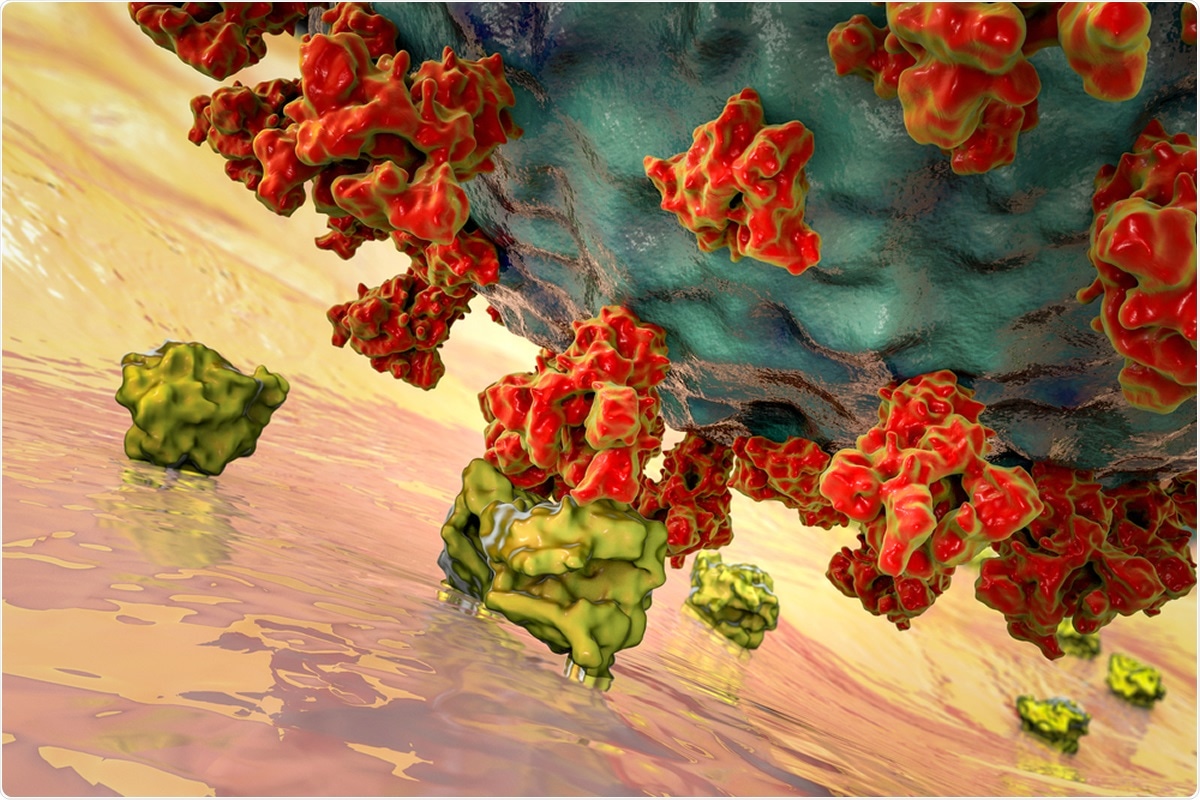
SARS-CoV-2 viruses binding to ACE-2 receptors on a human cell. SARS-CoV-2 uses clathrin-mediated endocytosis to gain access into cells. Image Credit: Kateryna Kon / Shutterstock

 This news article was a review of a preliminary scientific report that had not undergone peer-review at the time of publication. Since its initial publication, the scientific report has now been peer reviewed and accepted for publication in a Scientific Journal. Links to the preliminary and peer-reviewed reports are available in the Sources section at the bottom of this article. View Sources
ACE2 is a protein on the surface of many cell types. It is an enzyme that generates small proteins – by cutting up the larger protein angiotensinogen – that then go on to regulate functions in the cell.
Using the spike-like protein on its surface, the SARS-CoV-2 virus binds to ACE2 – like a key being inserted into a lock – prior to entry and infection of cells. Hence, ACE2 acts as a cellular doorway – a receptor – for the virus that causes COVID-19 disease.
The researchers added that this membrane system of the host cells or eukaryotic cells is an important cellular defense against invading viruses and microbes. However, the viruses tend to gain access into the cells by bypassing this complex mechanism. Antiviral drugs target this level of cellular entry of the virus, they wrote.
Infection rates of the virus
The team wrote that the numbers of confirmed CVOD-19 cases and the real number of people infected might be different. They said that antibody tests have revealed that 3 to 20 percent of certain communities have already been infected by the virus, indicating its high rates of infectiousness. These rates of infection in the community are far below those required for herd immunity, the researchers added.
How does the virus enter the cells? What is known?
The team of researchers explained that the virus has a transmembrane spike (S) glycoprotein that is capable of forming “homotrimers.” The S protein has S1 and S2 subdomains. Here the S1 codes for the receptor-binding domain that allows the virus to bind to the host cells and the S2 subdomain have a transmembrane domain that allows the virus membrane to fuse with the host cell membrane. The receptor on which the S glycoprotein binds is the angiotensin-converting enzyme 2 (ACE2) that is present on the host cells. Once the S protein binds to the ACE2, it is broken into S1 and S2 by “type II transmembrane serine protease TMPRSS2”, a cleaving enzyme. Another enzyme furin activates the S2 subdomain that allows fusion of the viral and cellular membrane and entry of the viral RNA.

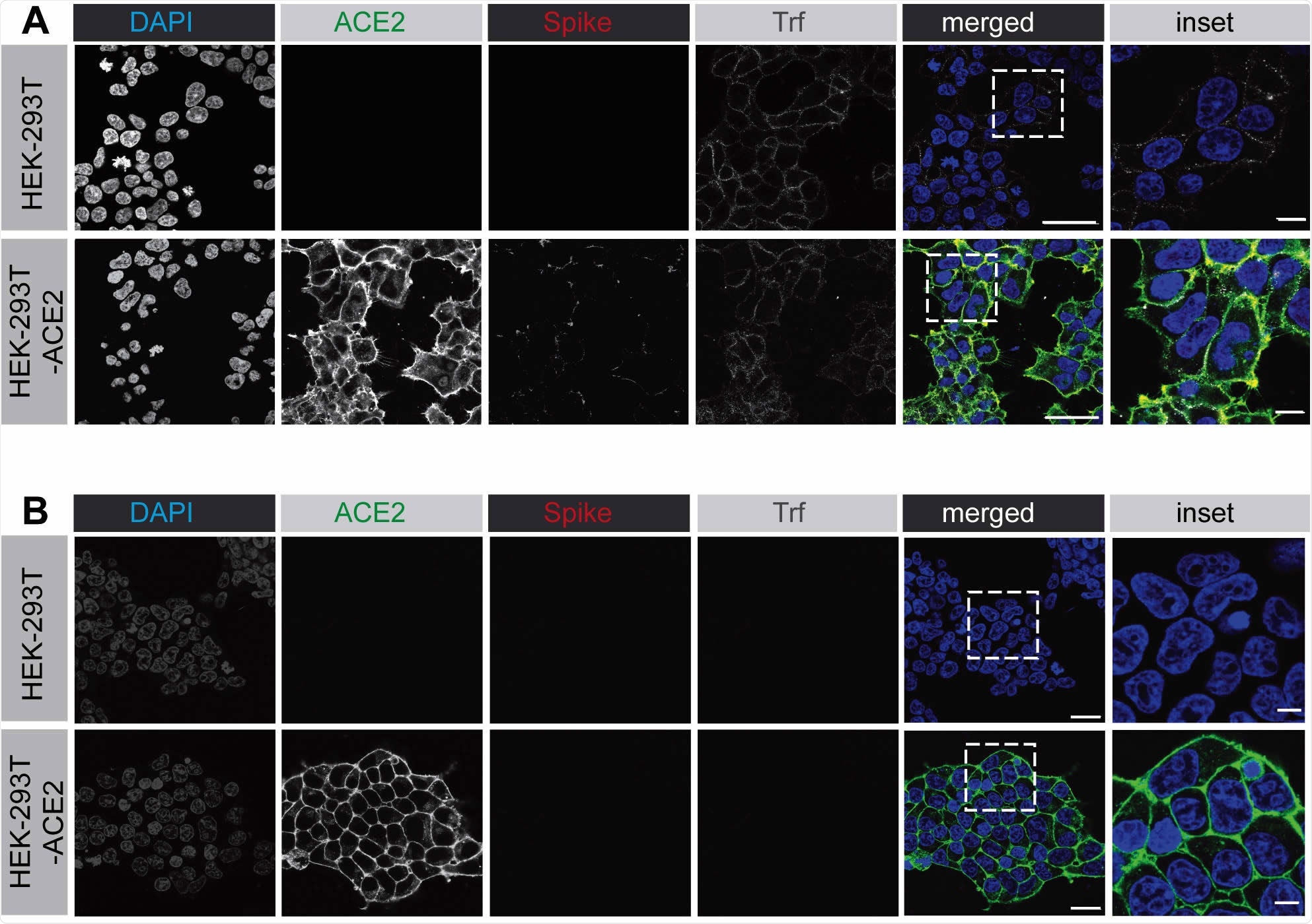
SARS-CoV-2 spike protein binds to the surface of HEK-293 cells expressing ACE2. (A) HEK-293 cells, wild-type (top row of images) or stably expressing ACE2 (bottom row of images) were incubated with purified, His6-tagged spike protein and with alexa-647 labelled transferrin for 30 min at 4°C. Following PBS wash, the cells were fixed and stained with DAPI to reveal nuclei, with an antibody selectively recognizing ACE2, and with an antibody recognizing the His6 epitope tag of the spike protein. Scale bars = 40 µm for the low mag images and 10 µm for the higher mag inset of the composite. (B) Experiment performed as in A except that the HEK-293 cells were briefly acid washed prior to fixation. Scale bars = 40 µm for the low mag images and 10 µm for the higher mag inset of the composite.
Endocytosis
The coat proteins of the virus thus do not enter the host cell. What enters is viral RNA. Another theory is that the ACE2/SARS-CoV-2 bound complex on the cell membrane is engulfed as a whole by the cell membrane by the process of endocytosis. This endocytosis includes capsid proteins.
Two methods of endocytosis of the whole virus/ACE2 receptor complex have been reported in the scientific literature. These are;
- Clathrin-mediated endocytosis (CME)
- Clathrin-independent process
What was done and what did the team find?
For this study, the team first took purified spike glycoprotein protein. They used lentivirus pseudotyped with spike glycoprotein. The whole simulation showed that the SARS-CoV-2 could undergo rapid endocytosis in the cell membrane after binding to the receptors.
They then used specific chemical inhibitors to stop each of the steps. The results revealed that this entry of the virus and receptor complex is through clathrin-mediated endocytosis. The team wrote, “Thus, it appears that SARS-CoV-2 first engages the plasma membrane, then rapidly enters the lumen of the endosomal system, strongly suggesting that fusion of the viral membrane occurs with the lumenal membrane of endosomes.”
Conclusions and implications
The team explained that Chloroquine (CQ) and hydroxychloroquine (HCQ) – antimalarial drugs that were initially thought to be successful in treating COVID-19 cases are known to clock this clathrin mediated endocytosis. Chlorpromazine, a drug used in psychiatric illness, is also known to disrupt this process of endocytosis the team wrote. The researchers wrote that their finding is hugely significant because if chemical inhibitors to this clathrin mediated endocytosis could be developed, a possible drug to prevent the SARS-CoV-2 infection could be developed. They said that clathrin-mediated endocytosis “should be considered as a key cellular pathway in any future drug target screens” for COVID-19.

 This news article was a review of a preliminary scientific report that had not undergone peer-review at the time of publication. Since its initial publication, the scientific report has now been peer reviewed and accepted for publication in a Scientific Journal. Links to the preliminary and peer-reviewed reports are available in the Sources section at the bottom of this article. View Sources