As coronavirus disease (COVID-19) continues to impact the world, an adequate understanding of SARS-CoV-2 replication and virulence properties may unveil potential pathways to disrupt the disease. Most of the attention thus far has been directed towards the spike glycoprotein responsible for receptor binding and cell entry.
In short, after the angiotensin-converting enzyme 2 (ACE2) receptor is recognized, the spike glycoprotein gets cleaved at two sites (S1/S2 and the S2' site) in order to facilitate viral entry into the cell. Accordingly, many researchers have focused their attention on a possibly critical insertion of a furin cleavage site found upstream of the S1 cleavage site in spike glycoprotein.
Kindred furin cleavage sites have been observed in other virulent pathogens like avian influenza, human immunodeficiency virus (HIV), and Ebola. More importantly, furin cleavage sites can be found in a myriad of other coronavirus family members – including MERS-CoV. Therefore, the acquisition of the furin cleavage site can be observed as a 'gain of function', possibly opening the door for the virus to jump to humans.
So the pertinent question is whether the furin cleavage site plays a significant role in SARS-CoV-2 infectivity and pathogenesis. This was recently tackled by a group of researchers from the University of Texas Medical Branch, Emory University, Icahn School of Medicine at Mount Sinai, University of Texas Health Science Center at Houston, and Bowie State University in the United States.

 This news article was a review of a preliminary scientific report that had not undergone peer-review at the time of publication. Since its initial publication, the scientific report has now been peer reviewed and accepted for publication in a Scientific Journal. Links to the preliminary and peer-reviewed reports are available in the Sources section at the bottom of this article. View Sources
Designing and appraising a mutant virus
In this manuscript, the scientists basically employed a reverse genetic system to generate a SARS-CoV-2 mutant that lacked the furin cleavage site insertion. More specifically, by utilizing a reverse genetic system for the SARS-CoV-2 WA1 isolate (obtained initially from the US CDC), they have generated a mutant virus that deleted the four amino acid insertion (ΔPRRA).
Vero E6 cells (derived from kidney epithelial cells of the African green monkey) or Calu3 cells (derived from non-small-cell lung cancer cell line that shows epithelial morphology and adherent growth) were infected with either wild type or ΔPRRA mutant viruses. Various methods subsequently ascertained relative viral protein levels.
The researchers next evaluated the fitness of the ΔPRRA mutant relative to wild type SARS-CoV-2 in a competition assay. More specifically, by utilizing plaque-forming units to determine the input, they have mixed the wild type and mutant viruses at different ratios in Vero E6 cells, subsequently evaluating their overall fitness with a reverse transcription PCR approach.
For in vivo studies, hamsters were challenged with the wild type or mutant virus by intranasal inoculation and then observed daily for the development of the clinical disease. The study also included a wide array of advanced genetic methods, such as deep sequencing analysis, phylogenetic tree building, the usage of sequence identity heat maps, and structural modeling.

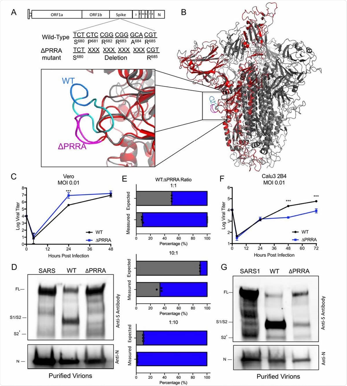
Distinct replication, spike cleavage, and competition 565 for ΔPRRA. A) Generation of a SARS-CoV-2 mutant deleting the furin cleavage site insertion from the spike protein. B) Structure of the SARS-CoV-2 spike trimer with a focus on the furin cleavage site (inset). Modeled using the SARS-CoV-1 trimer structure (PDB 6ACD) (14), the WT SARS-CoV-2 trimer (grey) with SARS-CoV-2 PRRA deletion mutant monomer overlay (red). The loop (inset), which is unresolved on SARS-CoV-2 structures (AA 691-702), is shown in cyan on SARS-CoV-2 with the PRRA sequence in blue. The loop region in the PRRA deletion mutant is shown in pink. C) Viral titer from Vero E6 cells infected with WT SARS-CoV-2 (black) or ΔPRRA (blue) at MOI 0.01 (N=3). D) Purified SARS-CoV, SARS-CoV-2 WT, and ΔPRRA virions were probed with anti-spike or anti-nucleocapsid antibody. 574 Full length (FL), S1/S2 cleavage form, and S2’ annotated. E) Competition assay between SARS-CoV-2 WT (black) and ΔPRRA (blue) showing RNA percentage based on quantitative RT-PCR at 50:50, 90:10, 10:90, 99:1, and 1:99 WT/ ΔPRRA ratio (N=3 per group). F) Viral titer from Calu3 2B4 cells infected with WT SARS-CoV-2 (black) or ΔPRRA (blue) at MOI 0.01 (N=3). G) Purified SARS-CoV, SARS-CoV-2 WT, and ΔPRRA virions were probed with anti-spike or anti-nucleocapsid antibody. Full length (FL), S1/S2 cleavage form, and S2’ annotated. P-values based on Student T-test and are marked as indicated: *<0.05 ***<0.001.
The role of furin cleavage site motif
In short, the loss of the furin cleavage site in the SARS-CoV-2 spike had a significant effect on infection and pathogenesis. The ΔPRRA mutant had augmented replication properties and improved fitness in Vero E6 cells in comparison to wild type SARS-CoV-2, as well as reduced spike processing. Conversely, the ΔPRRA mutant was attenuated in Calu3 cells and exhibited altered spike glycoprotein processing when compared to Vero cells.
Furthermore, plaque reduction neutralization tests that were conducted with sera from COVID-19 patients and monoclonal antibodies against the receptor-binding domain of the spike glycoprotein found a significant shift, with the mutant virus prompting consistently reduced neutralization titers.
In hamsters, the loss of the furin site attenuates SARS-CoV-2 induced disease but does not ablate the replication if the ΔPRRA virus. However, despite attenuated disease on primary infection, the ΔPRRA infected hamsters were actually protected from subsequent challenge with wild type SARS-CoV-2 – indicating the generation of a robust immune response.
The significance of the findings
"Overall, the data presented in this manuscript illustrate the critical role the furin cleavage site insertion in the spike protein plays in SARS-CoV-2 infection and pathogenesis", study authors confirm their findings in the bioRxiv preprint paper.
Biologically speaking, the loss of the furin site shifts the processing of the spike glycoprotein in a cell type-dependent manner. Moreover, the mutant ΔPRRA virus is impaired in its ability to replicate in certain cell types and to cause disease in vivo. Still, the results are complicated by observed augmented replication and fitness in Vero cells.
Finally, altered antibody neutralization profiles reveal a critical need to survey this mutation in the analysis of SARS-CoV-2 treatment options and vaccine candidates going forward. Also, understanding the details of the furin cleavage site may even help us in preventing potential future pandemics caused by coronaviruses.

 This news article was a review of a preliminary scientific report that had not undergone peer-review at the time of publication. Since its initial publication, the scientific report has now been peer reviewed and accepted for publication in a Scientific Journal. Links to the preliminary and peer-reviewed reports are available in the Sources section at the bottom of this article. View Sources
Journal references:
- Preliminary scientific report.
Johnson, B.A. et al. (2020). Furin Cleavage Site Is Key to SARS-CoV-2 Pathogenesis. bioRxiv. https://doi.org/10.1101/2020.08.26.268854.
- Peer reviewed and published scientific report.
Johnson, Bryan A., Xuping Xie, Adam L. Bailey, Birte Kalveram, Kumari G. Lokugamage, Antonio Muruato, Jing Zou, et al. 2021. “Loss of Furin Cleavage Site Attenuates SARS-CoV-2 Pathogenesis.” Nature, January, 1–10. https://doi.org/10.1038/s41586-021-03237-4. https://www.nature.com/articles/s41586-021-03237-4.