Using a circular polymerase chain reaction, researchers have developed a method for making severe acute respiratory syndrome coronavirus 2 (SARS-CoV-2) recombinant virus, with the ability to introduce mutations. The research by Japanese scientists is published on the preprint server bioRxiv* in September 2020.
As the COVID-19 pandemic continues to infect more and more people, there is a great need to understand the mechanisms by which the SARS-CoV-2 virus causes COVID-19 disease, infects, and replicates in host cells. To better understand how the disease develops, it is necessary to have a simple reverse genetics system for the virus.
Such systems are usually made using infectious clones having the entire viral cDNA. However, cDNA for coronaviruses are not available as the viral genomes are large. Instead, bacterial artificial chromosomes (BAC) or fragments of cDNA are joined together in vitro.
But, these systems have their disadvantages. In BAC systems, there may be undesired mutations caused by bacterial amplification, which then requires checking the entire genome every time, a time-consuming process. Joining together cDNA fragments is a complicated process requiring expertise.
Another method uses the circular polymerase chain reaction (CPER), to generate infectious clones of flaviviruses. cDNA fragments and a linker fragment are amplified by polymerase chain reaction (PCR). The amplified fragments are designed to include overlapping ends with other fragments, and hence can be extended as a circular viral genome. This viral genome is then introduced into the appropriate cells, and infectious viral clones are recovered.

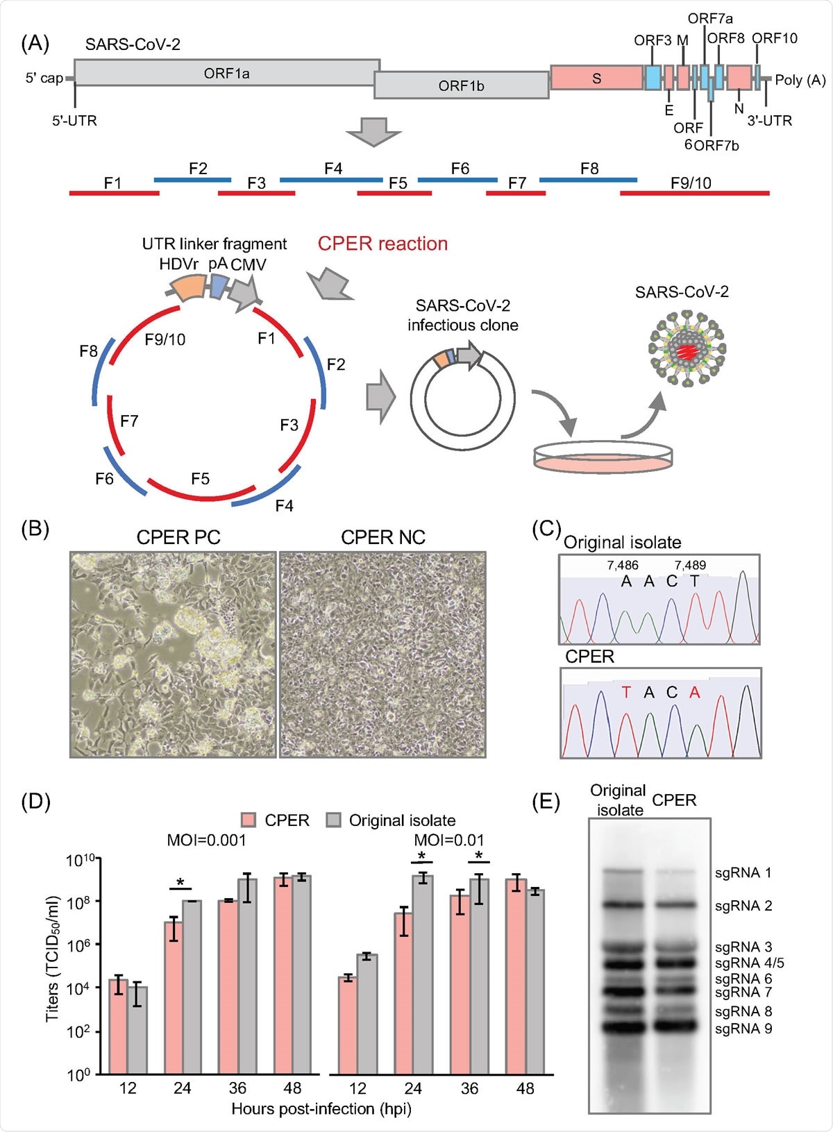
Establishment of CPER-based reverse genetics for SARS-CoV-2. (A) Schematic representation of a CPER approach for the generation of recombinant SARS-CoV-2. A total of 9 fragments (F1to F8, and F9/10) covering the full-length of the SARS-CoV-2 genome were amplified, then assembled with a UTR linker fragment including the HDVr, the BGH polyA signal and the CMV promoter by CPER. The resulting CPER products were transfected into the susceptible cells. (B) HEK293-3P6C33 cells were transfected with the CPER product and the bright field image was acquired at 7 days post-transfection (dpt) (left). As a negative control, the CPER product obtained without fragment F9/10 was transfected into cells and the bright field image was obtained at 7 dpt (right). (C) Genetic markers (2 silent mutations, A7,486T and T7,489A) in the recombinant SARS-CoV-2 genome. (D) Comparison of the growth kinetics of the recombinant SARS-CoV-2 generated by CPER with that of the original isolate. VeroE6/TMPRSS2 cells were infected with the viruses (MOI=0.001 or 0.01), and infectious titers in the culture supernatants of the SARS-CoV-2-infected cells were determined by TCID50 assay from 12 to 48 hours post-infection (hpi). (E) Northern blot analyses of subgenomic RNAs. RNAs extracted from cells infected with the parental virus and the recombinant SARS-CoV-2 recovered by CPER were subjected to Northern blot analyses.

 This news article was a review of a preliminary scientific report that had not undergone peer-review at the time of publication. Since its initial publication, the scientific report has now been peer reviewed and accepted for publication in a Scientific Journal. Links to the preliminary and peer-reviewed reports are available in the Sources section at the bottom of this article. View Sources
CPER for SARS-CoV-2
In a new study, a team of researchers used the CPER approach to generate infectious clones for SARS-CoV-2.
First, to test if the CPER approach could be used for SARS-CoV-2, the researchers used 10 viral gene fragments, which covered the entire viral genome, a linker fragment, along with a promoter to clone into a plasmid. Then, they used 10 cDNA fragments of the viral gene fragments, connected the 9th and 10th fragments by PCR, and performed CPER, when the full-length cDNA was clone obtained.
To check if the obtained products were viable, they infected cells with these products and studied the cells for changes because of the viral infection. They found recombinant viral genes formed after 7 days of transfection into the cells, but only in one type of cells, HEK293-3P6C33. This suggests that the transfection efficiency of the CPER products and the replication efficiency of the virus are important to form a recombinant virus.
The authors performed genetic sequencing of the viruses formed by the CPER method, which showed there was no contamination of the virus, and it maintained the genetic markers. They found only one difference in the genetic markers of the P0 virus, showing the reverse genetics system for the SARS-CoV-2 had high accuracy.
After testing several primer sets, the authors found the design of primers is important for efficient assembly of the circular genome. Hence, “further investigation of the CPER conditions (i.e., promoter sequences and primer sets) for SARS-CoV-2 may enhance the recovery of recombinants,” write the authors.
The researchers also investigated the kinetics of the recombinant virus compared to the parent SARS-CoV-2 virus. They found that the propagation of the recombinant virus was slower than that of the parent virus, but reached the levels similar to that of the parent virus.
Northern blot analysis showed eight subgenomic RNAs, both in the recombinant virus and the parent virus. Thus, the biological characteristics of the virus formed by the CPER method are similar to that of the parent virus.
Adding mutations
The researchers tested the CPER method to add reporter genes to the recombinant virus. They replaced a sequence of nucleotides in the viral genome by the sfGFP gene.
Upon sequence analysis after CPER and testing GFP fluorescence after expressing the fluorescent protein in cells, they found the recombinant virus had the added mutation. However, the growth rate of the mutated virus was slower than that of the parent.
However, when the researchers used NanoBiT, a split reporter having two subunits, high-affinity NanoBiT (HiBiT) and large NanoBiT (LgBiT), the growth kinetics was similar to that of the wild-type virus.
In their test, they inserted HiBiT luciferase gene and a linker sequence into the SARS-CoV-2 virus by PCR and then used CPER to generate recombinant SARS-CoV-2 with HiBiT. Previous studies have shown the HiBiT gene may be useful for animal experiments and drug screening.
Furthermore, the authors also showed they could insert a mutation in the spike protein of the virus, which has been seen in Europe and the Americas, without any change in the growth kinetics of the recombinant virus.
Thus, the authors say the CPER method can be a fast and straightforward tool for developing safe live-attenuated vaccines and characterizing mutations in the virus upon using vaccines or drugs.

 This news article was a review of a preliminary scientific report that had not undergone peer-review at the time of publication. Since its initial publication, the scientific report has now been peer reviewed and accepted for publication in a Scientific Journal. Links to the preliminary and peer-reviewed reports are available in the Sources section at the bottom of this article. View Sources
Journal references:
- Preliminary scientific report.
Torii, S. et al. (2020) Establishment of a reverse genetics system for SARS-CoV-2 using circular polymerase extension reaction. bioRvix. https://doi.org/10.1101/2020.09.23.309849
- Peer reviewed and published scientific report.
Torii, Shiho, Chikako Ono, Rigel Suzuki, Yuhei Morioka, Itsuki Anzai, Yuzy Fauzyah, Yusuke Maeda, Wataru Kamitani, Takasuke Fukuhara, and Yoshiharu Matsuura. 2021. “Establishment of a Reverse Genetics System for SARS-CoV-2 Using Circular Polymerase Extension Reaction.” Cell Reports 35 (3): 109014. https://doi.org/10.1016/j.celrep.2021.109014. https://www.cell.com/cell-reports/fulltext/S2211-1247(21)00328-4.