Akin to a myriad of other enveloped virus glycoproteins, SARS-CoV-2 spike glycoprotein is synthesized as a precursor that is subsequently cleaved in order to be able to exert fusion activity with the human cell membrane.
Depending on the exact sequence of spike glycoprotein at the S1/S2 junction, this cleavage occurs during the trafficking of the spike in the producer cell – either by host furin-like enzymes or by serine proteases at the cell surface (such as the transmembrane protease serine 2 or TMPRSS2).
The presence of a furin cleavage site at the S1/S2 junction is not unusual in human coronaviruses. More specifically, two of the four seasonal coronaviruses that are renowned for their efficient transmission in humans – hCoV-HKU1 and hCoV-OC43 – both contain furin cleavage sites. In contrast, MERS-CoV contains a suboptimal dibasic furin cleavage site.
On the other hand, the other two human seasonal coronaviruses (hCoV-229E and hCoV-NL63) do not contain furin cleavage sites in their spike glycoproteins, apparently without any loss of transmissibility. Hence, furin-mediated cleavage of spike glycoprotein is not an absolute prerogative for effective respiratory spread among humans.
So what is the situation with SARS-CoV-2? The insertion of four amino acids in its spike glycoprotein resulted in a suboptimal furin cleavage site (CS). However, a research group led by Dr. Thomas P. Peacock from the Imperial College London in the UK proposed a mechanism by which type of furin cleavage site is actually an advantage to the virus in the human airway, enabling successful human-to-human transmission.

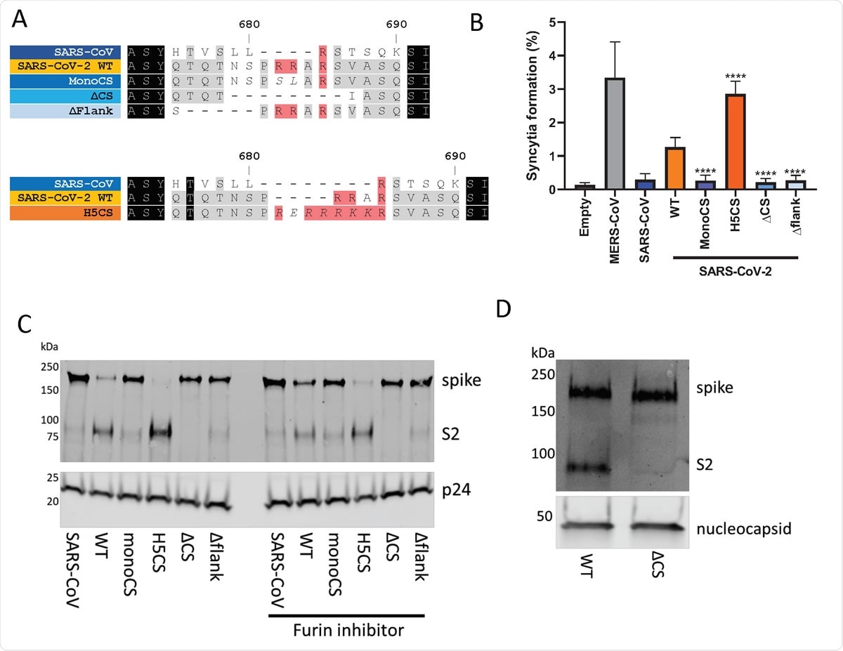
The SARS-CoV-2 spike contains a suboptimal polybasic furin cleavage site at the S1/S2 site. (A) Amino acid sequence alignment of coronavirus furin cleavage site mutants used in this study. Mutants with potential S1/S2 furin cleavage sites shown in shades of orange while mutants without furin cleavage sites shown in shades of blue. (B) Syncytia formation due to overexpression of different coronavirus spike proteins in Vero E6 cells. The percentage indicates the proportion of nuclei in each field which have formed clear syncytia. Statistical significance determined by one-way ANOVA with multiple comparisons against SARS-CoV-2 WT. **** indicates P value < 0.0001. (C) Western blot analysis of concentrated lentiviral pseudotypes with different coronavirus spike proteins. Levels of lentiviral p24 antigen shown as a loading control. Lentiviral pseudotypes labelled ‘furin inhibitor’ were generated in the presence of 5 µM Decanoyl-RVKR766 CMK, added 3 hours post-transfection. (D) Western blot analysis of concentrated WT and ΔCS SARS-CoV-2 viruses. Levels of nucleocapsid (N) protein shown as loading control.

 This news article was a review of a preliminary scientific report that had not undergone peer-review at the time of publication. Since its initial publication, the scientific report has now been peer reviewed and accepted for publication in a Scientific Journal. Links to the preliminary and peer-reviewed reports are available in the Sources section at the bottom of this article. View Sources
Generating spike mutants
In this study, the researchers used a combination of lentiviral SARS-CoV-2 pseudotypes harboring spike glycoprotein cleavage site mutations, as well as Vero passaged SARS-CoV-2 virus variants in order to investigate the molecular mechanism by which the polybasic cleavage site of the virus facilitates organized and efficient entry into lung cells.
More specifically, the significance of the spike glycoprotein polybasic cleavage site of SARS-CoV-2 was studied by generating a number of spike mutants that were predicted to regulate the efficiency of furin cleavage and testing them in cell cultures and laboratory animals (ferrets).
All human samples used in this extensive research project were obtained from the Imperial College Healthcare Tissue Bank. Finally, the ability of ferret sera to neutralize wild type SARS-CoV-2 virus was appraised by using a neutralization assay on the Vero E6 cell line (i.e., kidney epithelial-derived cells commonly employed for viral propagation).
Deciphering efficient viral transmission
"We show that pre-cleavage of the spike during viral egress enhanced entry of progeny virions into TMPRSS2-expressing cells such as those abundant in respiratory tissue", study authors explain their main study finding.
In other words, the polybasic insertion to the S1/S2 cleavage site gives SARS-CoV-2 a significant fitness advantage in TMPRSS2 expressing cells, which is likely a key prerequisite for efficient transmission of the virus between humans.
This was confirmed in animal studies where, in contrast with wild type SARS-CoV-2, a virus with a deleted furin cleavage site did not replicate to high titers in the upper respiratory tract of ferrets, and also did not transmit to cohoused sentinel animals (which is completely in agreement with similar experiments conducted in hamsters).
Proposing novel treatment options
These results unveil TMPRSS2 as a potential target for drugs and novel therapeutics. And while inhibition of TMPRSS2 protease activity does not prevent other infection paths that can occur via the endosome, this protease is indispensable to viral replication in airway cells.
"We have shown in this study that the protease inhibitor, camostat, is highly efficient at blocking SARS-CoV-2 replication in human airway cells, and we note that clinical trials are ongoing", study authors emphasize implications of their bioRxiv paper.
This study also has methodological value, as it confirms the impediments of relying on the Vero E6 cell line as a path towards developing drug classes that serve as entry inhibitors since they do not accurately mirror the favored entry mechanism of SARS-CoV-2 into human airway cells.
In any case, the authors conclude the paper by suggesting that a furin cleavage site in the SARS lineage of viruses is a cause of concern. Therefore, monitoring wild coronaviruses is a pivotal step in predicting and intercepting potential future pandemics.

 This news article was a review of a preliminary scientific report that had not undergone peer-review at the time of publication. Since its initial publication, the scientific report has now been peer reviewed and accepted for publication in a Scientific Journal. Links to the preliminary and peer-reviewed reports are available in the Sources section at the bottom of this article. View Sources
Journal references:
- Preliminary scientific report.
Peacock, T.P. et al. (2020). The furin cleavage site of SARS-CoV-2 spike protein is a key determinant for transmission due to enhanced replication in airway cells. bioRxiv. https://doi.org/10.1101/2020.09.30.318311.
- Peer reviewed and published scientific report.
Peacock, Thomas P., Daniel H. Goldhill, Jie Zhou, Laury Baillon, Rebecca Frise, Olivia C. Swann, Ruthiran Kugathasan, et al. 2021. “The Furin Cleavage Site in the SARS-CoV-2 Spike Protein Is Required for Transmission in Ferrets.” Nature Microbiology, April, 1–11. https://doi.org/10.1038/s41564-021-00908-w. https://www.nature.com/articles/s41564-021-00908-w.