An allosteric center or site is one where a molecule other than the substrate to the protein can bind, altering the conformation of the protein. A paper recently uploaded to the preprint server bioRxiv* by researchers at Chapman University in California (Feb 22nd, 2021) utilized computational methods to screen severe acute respiratory syndrome coronavirus 2 (SARS-CoV-2) mutants for affinity with the angiotensin-converting enzyme 2 (ACE2) receptor and monoclonal antibodies REG10987 and REG10933. The analysis allowed the group to identify allosteric centers that mediate long-range communication in the protein. The group suggests that it is these centers that allow the spike protein of SARS-CoV-2 to act in such a versatile manner, modulating the response to antibodies while conserving ACE2 affinity.


 This news article was a review of a preliminary scientific report that had not undergone peer-review at the time of publication. Since its initial publication, the scientific report has now been peer reviewed and accepted for publication in a Scientific Journal. Links to the preliminary and peer-reviewed reports are available in the Sources section at the bottom of this article. View Sources
Which mutations alter affinity?
Deep mutational scanning approaches to the receptor-binding domain (RBD) of SARS-CoV-2 identified constraints in the conformation of the protein regarding its ability to interact with ACE2, though also highlighted that many mutations in the RBD are well-tolerated and do not significantly affect binding affinity. In fact, many modifications were found to improve binding with ACE2 either by directly increasing the affinity, as with N501F, N501T, and Q498Y mutations, or enhancing RBD expression was found for V367F and G502D mutations. Mutations to the ACE2 receptor can also have similarly enhancing effects with regards to binding with the RBD.
RBD mutations N439/R426, L452/K439, T470/N457, E484/P470, Q498/Y484, and N501/T487 were noted in particular to enhance binding affinity with ACE2. Several mutant strains of SARS-CoV-2, including the UK variant B.1.1.7, the South African variant B.1.351, and the P.1 (formerly B.1.1.28.1) strain identified in Tokyo each exhibit some or all of these mutations, and are noted for their higher transmissibility.
Mutations that were involved in escape from monoclonal antibodies REG10987 and REG10933 were shown to have only a minor negative impact on ACE2 binding and RBD folding. Mutation at the E484 site had the largest effect on antibody binding, which is in line with other studies that note a ten-fold loss in the affinity of the antibodies towards the RBD due to this change.
The group applied the same structural and functional studies to mRNA-vaccine induced antibodies to SARS-CoV-2 and the mutants of importance, importantly noting that the affinity of the antibodies towards the K417N/E484K/N501Y mutants was lower by a small but significant margin. Vaccine-induced immunity may, therefore, be compromised by alternate strains of SARS-CoV-2. These key mutations were also shown to lessen or eliminate the affinity of 14 of the 17 most-utilized monoclonal antibodies against the virus.

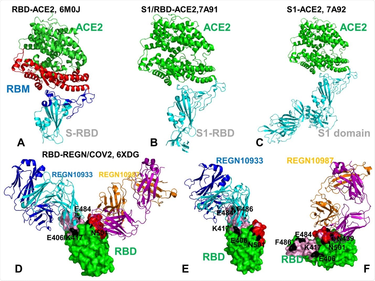
Crystal structures of the SARS-CoV-2 RBD and S1 domain complexes with ACE enzyme and REGN-COV2 antibody cocktail. (A) Structural overview of the SARS-CoV-2 RBD complex with ACE2 (pdb id 6M0J). The SARS-CoV RBD is shown in cyan ribbons and the RBM region is in blue ribbons. The subdomain I of human ACE2 is shown in red ribbons and the subdomain II is shown in green ribbons. The structure of ACE2 consists of the N-terminus subdomain I (residues 19-102, 290-397, and 417-430) and C-terminus subdomain II ( residues 103-289, 398-416, and 431-615) that form the opposite sides of the active site cleft. (B) The crystal structure of the dissociated S1 domain form in the complex with ACE2 (pdb id 7A91). S1-RBD is in cyan ribbons and ACE2 is in green ribbons. (C) The crystal structure of the fully dissociated S1 domain in the complex with ACE2 (pdb id 7A92). S1 domain of the SARSCoV- 2 S protein is in cyan ribbons and ACE2 is in green ribbons. (D) The cryo-EM structure of the SARS-CoV-2 RBD in the complex with REGN10933/REGN10987 antibody cocktail. The RBD region is shown in green surface. REGN10933 Fab fragment is shown in ribbons with heavy chain in cyan and light chain in blue. REGN10987 is in ribbons with heavy chain in orange and light chain in purple. The positions of functional residues targeted by mutational variants and antibody-escaping mutations are E406, K417, E484 and N501 are annotated and highlighted as black patches on the RBD surface. (E) A close-up of the SARS-CoV-2 RBD interactions with REGN10933. The RBD is shown in green surface. REGN10933 Fab fragment is shown in ribbons with heavy chain in cyan and light chain in blue. The REGN10933 antibody epitope on RBD is highlighted in cyan patches on the surface. The positions of E406, K417, E484, F486, N501 are shown as black surface patches on the RBD. (F) A close-up of the SARSCoV- 2 RBD interface with REGN10987. The red patches correspond to the REGN10987 epitope. The positions of E406, K417, N439, E484, F486, N501 are shown as black surface patches on the RBD.
How can ACE2 affinity increase while antibody affinity is decreased?
K417, E484, and N501 residues were found to be key interacting centers with a high degree of structural plasticity that allow mutants at these positions to improve binding affinity with ACE2. E406, N439, K417, and N501 residues act as allosteric centers that anchor together segments of the molecule to mediate long-range interactions. Mutations at these sites alter the protein structure and neutralize the ability of monoclonal antibodies to bind with the spike protein of SARS-CoV-2 while maintaining ACE2 affinity. These sites are at hinge centers within the spike protein, and are generally involved in collective movements of the molecule. For example, the RBD is constantly in motion, alternating through closed and open conformations. This explains the ability of these mutations to aid in antibody escape while maintaining or enhancing ACE2 affinity.
SARS-CoV-2 strains that are unable to bind with ACE2 are at a direct evolutionary disadvantage and are quickly eliminated from the population. Highly adaptable and plastic allosteric centers allow the collective motions of the protein to be disrupted, becoming immune to monoclonal antibodies that require a specific structural arrangement to interact. The static conformation of the ACE2-interacting face of the spike protein remains largely unchanged by these mutations, only the behavior of the protein arrangement.

 This news article was a review of a preliminary scientific report that had not undergone peer-review at the time of publication. Since its initial publication, the scientific report has now been peer reviewed and accepted for publication in a Scientific Journal. Links to the preliminary and peer-reviewed reports are available in the Sources section at the bottom of this article. View Sources
Journal references:
- Preliminary scientific report.
Comparative Perturbation-Based Modeling of the SARS-CoV-2 Spike Protein Binding with Host Receptor and Neutralizing Antibodies : Structurally Adaptable Allosteric Communication Hotspots Define Spike Sites Targeted by Global Circulating Mutations, Gennady Verkhivker, Steve Agajanian, Deniz Yazar Oztas, Grace Gupta bioRxiv 2021.02.21.432165; doi: https://doi.org/10.1101/2021.02.21.432165, https://www.biorxiv.org/content/10.1101/2021.02.21.432165v1
- Peer reviewed and published scientific report.
Verkhivker, Gennady M., Steve Agajanian, Deniz Yazar Oztas, and Grace Gupta. 2021. “Comparative Perturbation-Based Modeling of the SARS-CoV-2 Spike Protein Binding with Host Receptor and Neutralizing Antibodies: Structurally Adaptable Allosteric Communication Hotspots Define Spike Sites Targeted by Global Circulating Mutations.” Biochemistry 60 (19): 1459–84. https://doi.org/10.1021/acs.biochem.1c00139. https://pubs.acs.org/doi/10.1021/acs.biochem.1c00139.