Several pieces of evidence have suggested that blue light can inactivate various pathogenic bacteria. Previously in vitro and in vivo studies using blue light in the spectral range of 400–470 nm against Gram-positive, Gram-negative, fungi, and mycobacteria were carried out.
The antimicrobial activity of blue light is due to the absorption of these wavelengths by the porphyrins and other chromophores that are present within bacteria. This leads to the generation of singlet oxygen and other reactive oxygen species (ROS) that causes non-specific oxidative damage to vital structures and microbial inactivation.
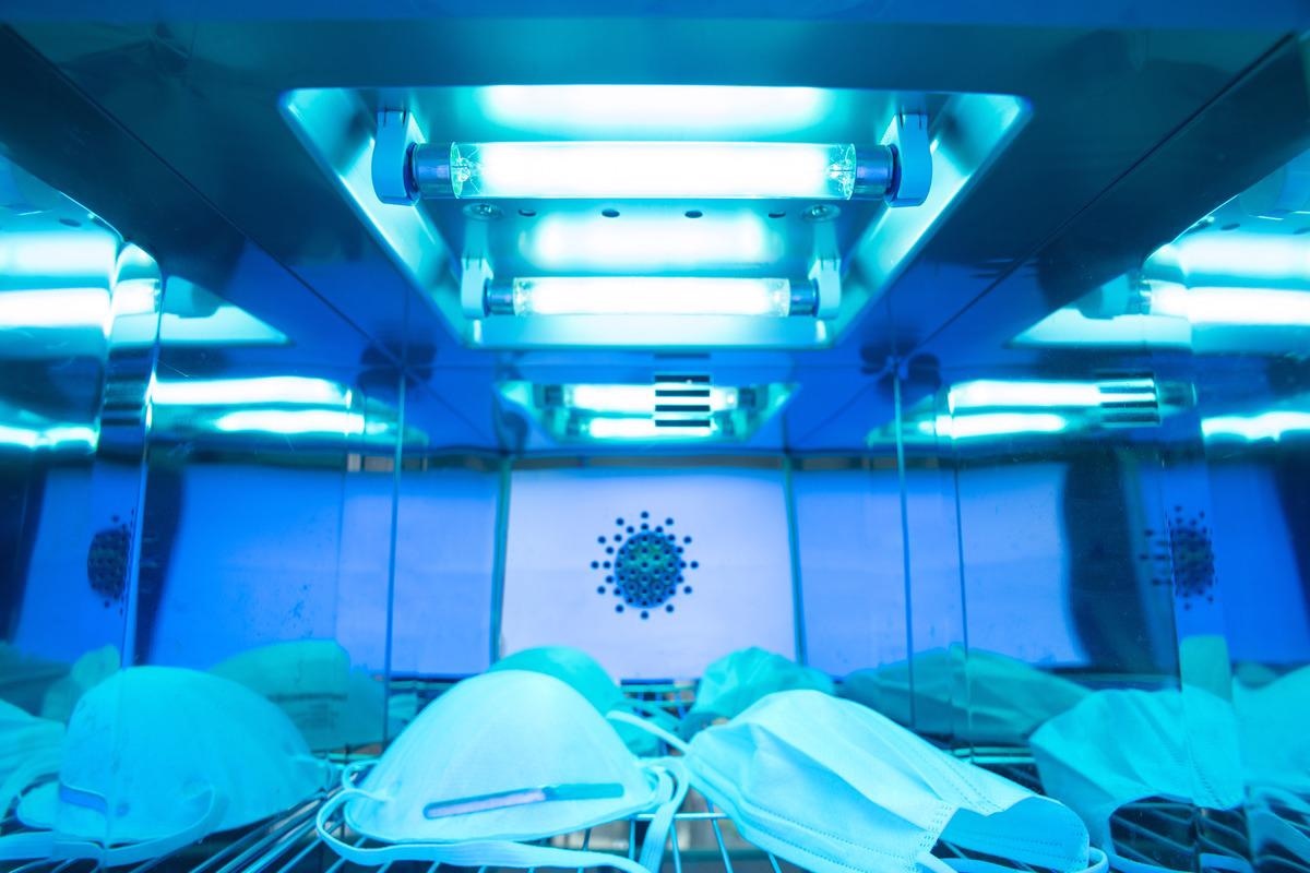
 Study: Efficient Inactivation of SARS-CoV-2 and Other RNA or DNA Viruses with Blue LED Light. Image Credit: Nor Gal/Shutterstock
However, the studies focusing on the virucidal activity of blue light are limited. One study indicated that bacteriophage/C31 was susceptible to high doses of blue light. Other studies indicate that high doses of blue light were effective against murine leukemia virus and feline calicivirus. However, further investigation is required as the mechanism of inactivation has not been determined yet.
A new study published in Pathogens analyzed the effect of blue light on severe acute respiratory syndrome coronavirus 2 (SARS-CoV-2), respiratory syncytial virus, and adenovirus. All three viruses can be transmitted via aerosol while SARS-CoV-2 was found to survive on surfaces for a long time.
The study
The study included a blue light LED instrument that emitted at 410–430 nm wavelength. All the three viruses were cultured in Vero E6 cells following which they were exposed to blue LED light for three, five, ten, or fifteen minutes at room temperature. A control was prepared separately which was not exposed to blue light.
Along with SARS-CoV-2, its trimeric spike protein was also exposed to blue LED light. Thereafter immunoblotting was carried out. Finally, the treated spike gene was amplified and protein analysis was done using fluorescence spectroscopy.
Findings
The results indicated that the exposure of the samples to blue LED light for 15 minutes was optimum as it led to complete inactivation of the viral load. However, the nucleic acids were not degraded, and no protein was denatured even at 15 minutes of exposure time.
Whether the presence of photosensitizers was necessary to activate LED in the case of SARS-CoV-2 was determined using swab samples from COVID-19 positive subject. The samples were placed in a physiological solution that did not contain any photosensitizer. Half of the sample was irradiated while the other half was kept at room temperature. It was found that the viral load in the irradiated sample was significantly reduced as compared to the untreated sample.
To determine whether substances present in the human respiratory droplets or biological samples could behave as photosensitizers, the SARS-CoV-2 spike protein suspended in sterile water or transport media was irradiated with blue LED light for 15 minutes. The fluorescence emission spectra of both the native and unfolded (incubated with a chaotropic agent) SARS-CoV-2 spike protein were obtained. After denaturation, a blue shift was observed in the case of the protein suspended in transport media as compared to sterile water.
Furthermore, the fluorescence spectra of the spike protein in transport media were analyzed before and after exposure to blue LED light. It was found that a significant reduction in fluorescence intensity occurs after exposure to blue LED light.
Conclusions
It can be concluded that the current study provided some possible explanations for the mechanisms by which antiviral activity is exerted by blue LED light. The study also indicated that the alteration of the SARS-CoV-2 spike protein required the presence of photosensitizers. Also, the development of low-cost light-based devices that can inactivate viruses can be of great utility in limiting their spread at hospitals, especially during the pandemic.