The coronavirus disease 2019 (COVID-19) pandemic has caused unprecedented morbidity and mortality globally. Despite the recovery of a large proportion of SARS-CoV-2-infected individuals, there are concerns regarding persistent injury in a few organs, such as the lungs, following COVID-19. However, data on the long-term pulmonary manifestations and function after 2.0 years of COVID-19 are limited.
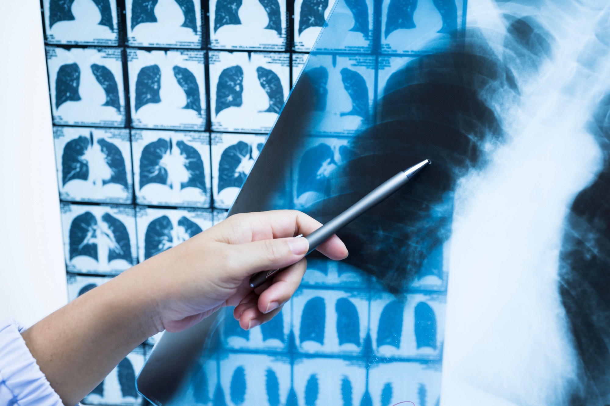
 Study: Longitudinal Assessment of Chest CT Findings and Pulmonary Function in Patients after COVID-19. Image Credit: Komsan Loonprom / Shutterstock
About the study
In the present prospective study, researchers assessed the long-term pulmonary sequelae of COVID-19.
The study comprised polymerase chain reaction (PCR)-confirmed SARS-CoV-2-positive individuals, aged between 18 and 80 years, who received hospital discharge following COVID-19 between 15 January and 10 March 2020, were eligible for the study. The team excluded individuals who did not undergo chest computed tomography scans at hospital admission, those with poor computed tomography scan quality, those with completely resolved pulmonary pathologies at hospital discharge, and those with a prior history of pulmonary diseases.
The team performed chest computed tomography investigations and lung function tests at six months (between 20 June and 31 August 2020), one year (between 20 December 2020 and 3 February 2021), and two years (between 16 November 2021 and 10 January 2022) following the onset of COVID-19 symptoms. Residual lung abnormalities such as interstitial lung abnormalities (ILAs) of the fibrotic type and the non-fibrotic type were analyzed during follow-up computed tomography investigations.
Additionally, the association between the residual pulmonary pathologies and pulmonary functional alterations was evaluated. Health records from the acute COVID-19 phase of all participants were reviewed. Three experienced radiologists independently analyzed the computed tomography scans, blinded to clinical data, and disagreements were resolved by consensus.
In-person interviews were conducted at the follow-up visits. At each visit, the participants underwent high-resolution CT scans and completed questionnaires on respiratory symptoms, including exertional dyspnea, cough, and expectoration. The team assigned CT scores according to the area of pulmonary lobe involvement with no involvement, <5.0% involvement, 5.0% to 25.0% involvement, 26.0% to 49.0% involvement, 50.0% to 75.0% involvement, and >75.0% involvement scores as 0.0,1.0,2.0,3.0,4.0, and 5.0, respectively. The individual scores for each lung lobe were summed up to calculate the total CT score.
Results
Out of 1,251 initially eligible individuals, 560 individuals without chest CT scans at hospital admission, 56 individuals with poor computed tomography scan quality, 294 individuals with entirely resolved pulmonary abnormities, and 95 individuals with a previous history of pulmonary disease were excluded from the analysis. In addition, 71 individuals who were unwilling to participate in the study and six who died due to non-COVID-19 reasons during follow-up were excluded.
As a result, 144 participants who had recovered from COVID-19 were followed up for 2.0 years, among whom the median participant age was 60 years, and most of them (n=79) were male. In addition, pulmonary function tests were completed by 110, 103, and 129 individuals at six months, one year, and two years post-COVID-19.
Most of the study participants (78%, n=112) suffered from severe SARS-CoV-2 infections, and the condition of 4.0% of them (n=six) was critical. Acute respiratory distress syndrome was documented in 19% (n=27) of the individuals. Among the study participants, over two years of SARS-CoV-2 infection, the incidence of ILAs gradually decreased (54.0%, 42.0%, and 39.0% at six months, one year, and two years, respectively).
Likewise, gradual reductions in the total CT scores with improved pulmonary function were observed. However, there were no statistically significant differences between the one-year and two-year follow-ups. The diffusing capacity of the lungs for carbon monoxide (DLco) values at six months, one year, and two years were 80, 82, and 84, respectively. DLco severity at the three follow-up visits did not show significant differences.
At two years of follow-up, the average total CT score was 2.0, with reticulation accounting for most pulmonary abnormalities. After two years of COVID-19 recovery, 39% (n=56) of participants showed ILAs, of which 23% (n=33) and 16% (n=23) were of the fibrotic type and non-fibrotic type, respectively, and the remaining 61% of participants (n=88) showed complete resolution.
Abnormal DLco (43% versus 20%) and pulmonary symptoms (34% versus 15%) were more prevalent among individuals with ILAs compared to individuals with complete resolution. Among individuals with ILAs, participants with ILAs of the fibrotic type more frequently experienced respiratory symptoms (45% versus 17%) and had lower DLco values (60% versus 22%) compared to individuals with ILAs of the non-fibrotic type.
CT scan findings indicating ILAs included reticular pathologies or ground-glass opacities (GGOs), traction-type bronchiectasis, lung architectural distortion, non-emphysematous lung cysts, and honeycomb-like patterns. Fibrotic interstitial lung abnormalities mainly comprised lung distortion, traction bronchiectasis, and honeycomb-like patterns, whereas the non-fibrotic interstitial lung abnormalities were mainly of the reticular or GGO type.
Overall, the study findings showed that two years following SARS-CoV-2 infection, more than one-third of the study participants had persistent ILAs associated with respiratory symptoms and decreased diffusion pulmonary function. The findings indicated that COVID-19 convalescents must be assessed longitudinally to identify and manage the long-term pulmonary complications of COVID-19.