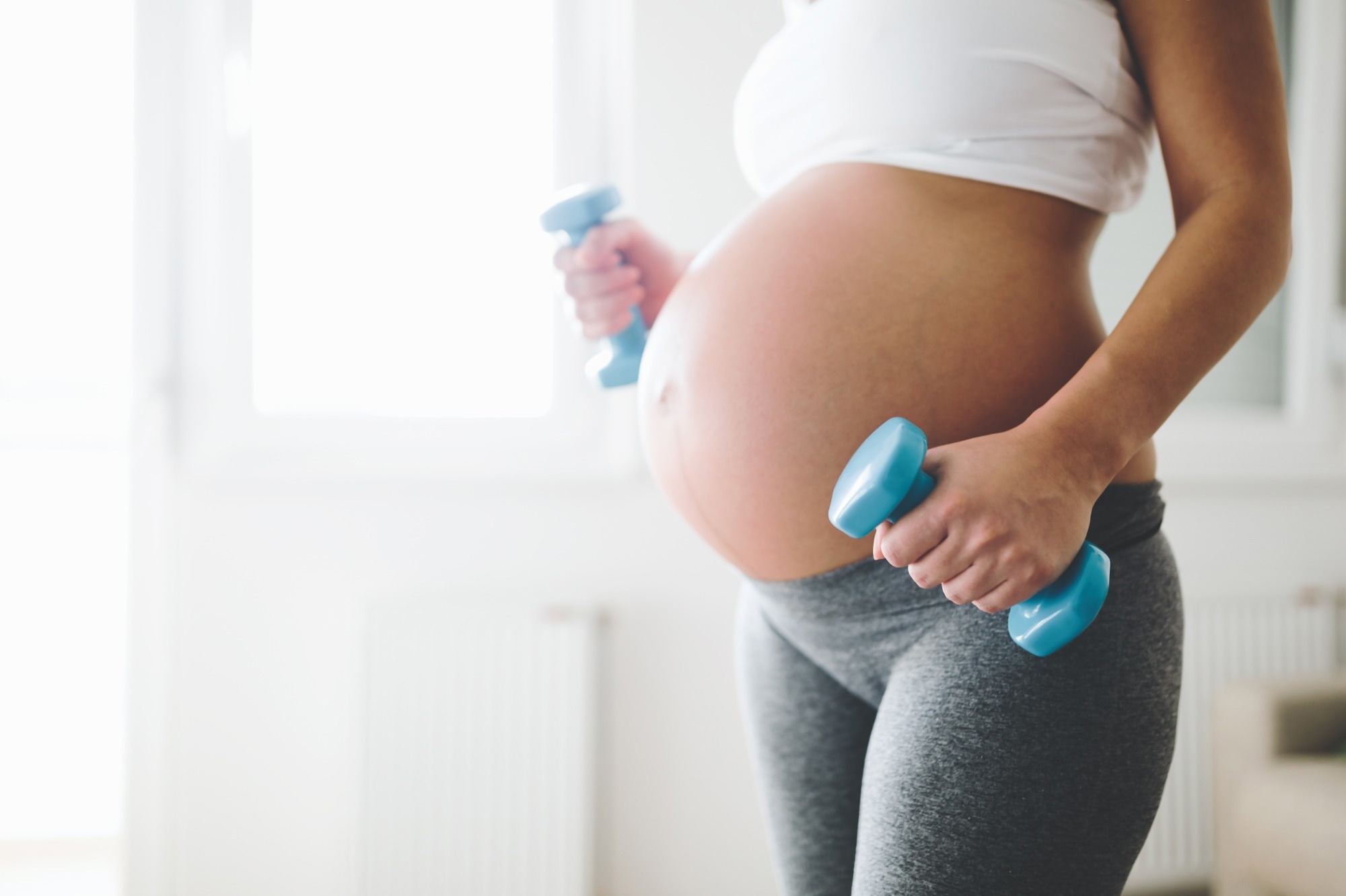
Exercise during pregnancy has been associated with a reduced risk of gestational diabetes and preeclampsia. According to recommendations from the American College of Obstetricians and Gynecologists, a healthy pregnant woman should exercise at moderate intensity for at least 150 minutes a week.
In the United States, about 1% of pregnancies result in complications due to cardiovascular disease (CVD), which could lead to maternal mortality and morbidity. Thus, it is important to understand the effect of moderate-intensity exercise in pregnant women with underlying cardiac conditions.
 Study: Moderate Intensity Exercise in Pregnant Patients with Cardiovascular Disease: A Pilot Study. Image Credit: NDAB Creativity / Shutterstock.com
Background
An abnormal umbilical artery Doppler test may indicate abnormal fetal and neonatal events, such as intrauterine growth restriction, which would require admission to the neonatal intensive care unit (NICU) once the infant is born. A very small number of studies have highlighted the importance of maternal exercise during pregnancy to achieve favorable fetal Doppler profiles.
One previous randomized-controlled trial assessed how exercise affected the biochemical markers associated with cardiac health in patients with gestational diabetes. This study reported an inverse association between exercise and circulating maternal C-reactive protein (CRP) levels.
About the study
A recent American Heart Journal study assessed the effect of moderate-intensity exercise in pregnant women with CVD. This prospective pilot study determined whether moderate intensity exercise is beneficial for pregnant women with pre-existing CVD based on umbilical artery Doppler ultrasound systolic to diastolic (S/D) ratio performed between 32-34 weeks of gestation.
The authors recruited participants from the Maternal Fetal Medicine Clinic at Brigham and Women’s Hospital in Boston, Massachusetts. The impact of exercise intensity and duration on hypertensive disorders, maternal weight gain, premature delivery, mode of delivery, gestational diabetes, birth weight, and neonatal Apgar scores were analyzed.
All participants were over 18 years of age and had a history of CVD; however, those with pulmonary hypertension, severe valvular heart disease, cyanosis, or pacemaker implantation were excluded from the study. In addition, individuals who experienced ectopic pregnancy were not included in the study cohort.
Participants were shown a ten-minute exercise video created by a certified exercise physiologist. The exercise was designed so that each exercise session would yield 60-70% of the age-predicted maximum heart rate or a suitable score on the exertion scale. Participants were asked to follow those exercises for 20 to 60 minutes for at least four days each week.
All women recruited in this study wore an activity tracker, which measured heart rate and step data throughout the pregnancy, particularly during exercise sessions.
Study findings
A total of 79 pregnant women satisfied all eligibility criteria and were included in this study. Among these, 37 had a history of CVD, and 42 were healthy participants.
Some participants from both groups did not complete the study, as they were not able to adhere to the exercise prescription or had issues with an activity tracker. Thus, the final cohort consisted of 24 participants in the CVD group and 28 in the control group.
Women belonging to the control group had greater exercise duration and intensity the rates of adverse maternal and fetal events remained the same between the groups. The overall outcomes in both groups were favorable. Marginally high NICU admission rates and smaller fetus size relative to their gestational age were observed in the CVD group.
Notably, no participants from either group developed gestational diabetes. In the future, more research is needed to study serum biomarker levels during pregnancy and the postpartum period.
Most of the study cohort exhibited decreased circulating CRP levels six weeks postpartum. However, 65% of the CVD group and 55% of the control group had CRP levels greater than 3 mg/L, which is associated with a high risk of future CVD.
Over 60% of participants in the CVD group had congenital heart disease, whereas four underwent repaired tetralogy of Fallot (TOF). Aside from a few NICU admissions, no significant difference in adverse maternal and neonatal outcomes was found between the two study groups.
The wearable activity data revealed a decrease in exercise intensity in the CVD group. This decrease was particularly evident during the third trimester.
Conclusions
The current study is the first to investigate the effect of exercise during pregnancy in women with a history of CVD. Exercise during pregnancy was found to be beneficial to maternal health, which has been associated with decreased postpartum weight gain, lower CRP levels, and reduced risk of gestational diabetes.
The study findings must be validated in the future using a large sample size. Furthermore, a tailored fitness program should be designed for pregnant women, considering their specific CVD conditions.