My name is Laura Kuett, and I recently graduated with a Ph.D. from the University of Zurich. My research was in breast cancer, specifically studying the tumor microenvironment. What drew me to this research was the method used; imaging mass cytometry.
This method is very computationally challenging; therefore, it was great to find out how it is used in breast cancer – one of the most common cancer types among women. I was excited to work in a field that affects so many women.
What is 3D Imaging Mass Cytometry, and what insights can it provide on human disease? What are the benefits of using this technique when analyzing tissues compared to other techniques available?
3D imaging mass cytometry is an extension of imaging mass cytometry, a method analogous to immunofluorescence. Similarly to immunofluorescence, this method uses antibodies to target proteins. While immunofluorescence uses a handful of targets, imaging mass cytometry can use up to 40 targets, and this is all thanks to metal tag antibodies.
In imaging mass cytometry, the tissue is stained with those metal tag antibodies, and then the tissue is ablated by laser to capture the counts of those metals in each tissue spot. These intensities are then translated into an image.
3D imaging mass cytometry is a workflow that I used during my Ph.D. It extends the imaging to the third dimension by carrying out a series of 2D images from tissue sections and then computationally reconstructing it into a 3D model.
Thanks to its multiplexity in having up to 40 different targets, this method really enables users to capture the different cell types in a tissue. For instance, it can capture different immune cell types but also different states of tumor cells, such as whether the cells are dividing fast, dying, or in different metabolic states.

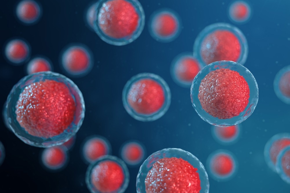
Image Credit: Rost9/Shutterstock.com
The important thing is that we can get the information of the different cell types together with the spatial information so that we can really study how biology happens in the tissues.
What are the benefits of using this technique when analyzing tissues compared to the other techniques that are available at the moment?
The main benefits of imaging mass cytometry are the high multiplexity, the ability to target 40 proteins simultaneously, and the spatial information we can get. This can provide great insights into tissue biology.
As a speaker at SLAS EU 2023, you gave a talk on the “Analysis of tissues at single-cell level with 3D Imaging Mass Cytometry”. Please can you give an overview of what you discussed in this talk?
In my talk, I gave an overview of the 3D imaging mass cytometric workflow. My main aim was to shine a light on the benefits of 3D imaging versus 2D imaging. 2D imaging is done very commonly across different fields, but I try to illustrate the benefits of 3D imaging, specifically multiplex single cell level 3D imaging, using breast cancer as an example.
I then used examples to show that 3D imaging enables us to intuitively and quickly capture the patterns that different cell types have in a tissue. 3D imaging can also capture phenomena that tend to go unnoticed with 2D imaging, such as potentially invasive tumor cells. With 3D imaging, we can see that those individual single cells are not attached to the bulk of the tumor.
One of the tracks of SLAS EU 2023 is Shaping the Future of Therapeutics. How do you believe 3D Imaging Mass Cytometry is shaping the future of therapeutics?
At the moment, I think 3D imaging mass cytometry is beneficial to basic research because it can give very detailed information about tissue biology and enable users to see and learn about tissues in more detail than ever before. I hope that translates into new hypotheses, eventually leading to new therapeutic targets.
Another SLAS EU track for this year is “Biology Unveiled.” How does the subsequent analysis of mass cytometry images unveil relevant biological information?
We live in a 3D world, and biology happens in 3D. Tissues are inherently 3D structures. Seeing the tissues visually in 3D and interacting with those 3D models would facilitate a greater understanding of tissue biology. I hope researchers can use these methods to learn more about tissue biology in health and disease.
Shaping the Future of Therapeutics with 3D Imaging Mass Cytometry
SLAS is an international professional society aiming to transform research by promoting collaboration at the intersection of science and technology. What recent technological advances that influenced the study of the tumor microenvironment at the single-cell level?
One of the biggest advances in recent times has been microfluidics. This has enabled us to separate individual single cells in a high-level manner and then carry out genome sequencing and transcriptome sequencing. This allows us to obtain a massive amount of information about the cells.
There have also been recent advances that have attempted to combine this with protein-level imaging methods. Combining multiplex imaging methods with single-cell data from the genome and transcriptome and building up this huge amount of information regarding tissues could enable us to learn more about biology in real depth.
I think conferences like SLAS, which have a very strong industry component, are great for researchers to see and learn what is needed to bring their research into action.
As a Ph.D. student in the field of oncology, how hopeful are you about the future of cancer research?
I am very hopeful about the future of cancer research, coming from the field of breast cancer. Breast cancer is a very well-studied cancer type because of the huge amount of funding that has gone into breast cancer research.

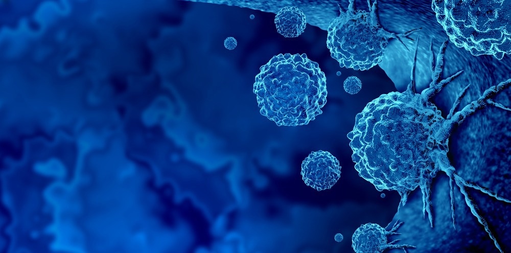
Image Credit: Lightspring/Shutterstock.com
I think this illustrates what can be achieved if a disease type is funded at such a high level. There have been incredible advancements in personalized therapies, disease prevention, and screening methods. I hope that other tumor types will have this level of funding and will have similar success in treatments in the future.
What is next for you and your work? Are you involved in any exciting upcoming projects?
I had a great Ph.D. experience. I worked with fantastic people, and I had an exciting project. Thanks to my Ph.D., I had a chance to work in a big scientific consortium that enabled collaboration with great scientists across the world.
But it was not only scientists but also game designers and animators because the whole big consortium project involved developing a virtual reality software for analyzing single-cell data. Game designers and animators helped to bring this to life.
This experience really showed me what is possible and where I could use my technical, scientific, communication, and teamwork skills. I have now transitioned into industry, and I work in a digital health startup in the field of chronic cough.
This startup combines technology, software development, and machine learning to improve people's lives with chronic coughs. I am really excited about this work.
About Laura Kuett
Laura completed her Ph.D. degree in computational cancer biology in 2022 from the University of Zurich. Her research at Bernd Bodenmiller's group focused on high-dimensional single-cell spatial tissue analysis with a specific interest in 3D imaging and metastatic processes in breast cancer. Before starting the Ph.D. degree, Laura studied molecular microbiology at the University of Aberdeen, Scotland, and undertook a one-year-long placement at the Luxembourg Centre of Systems Biomedicine, working on metabolic modeling of the gut microbiome. Since finishing her Ph.D., Laura works in a health technology start-up in Switzerland, focusing on regulatory processes and project management.