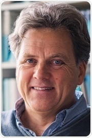
In this interview, News-Medical.net speaks to Prof. Klaus Gerwert of Ruhr University, about how histopathology problems can be solved using infrared microscopy.
What is histopathology and how is microscopy used in conjunction with histopathology to diagnose disease?
In classical histopathology, you take a biopsy, slice the biopsy in thin sections, and then you stain the biopsy with hematoxylin and eosin stain (H&E), a specific compound. This visualizes some morphological changes within the tissue. The pathologist then looks at these morphological changes and they can classify the disease and identify if there is cancer present or not. This is a very personally-driven decision. Laser-based microscopy is not only good to identify cancer quickly, but it's also very important to make a differential diagnosis.
The differential diagnosis is prognostic, and this is what the clinician needs for the therapeutic decision. They have to know which specific subclass a patient has. For example, in lung cancer, it's important not only to know if small lung canceris present, but it's also important to know whether it is adenocarcinoma, and if so, it's important to know which subclass of adenocarcinoma it is.
We have shown by using the quantum cascade laser (QCL) based microscope that you can do all of these differential diagnoses and that this is what the clinicians need in order to make a specific therapeutic decision. Now we need to look at sensitivity and specificity versus the new label-free digital pathology approach and compare it to the classical histopathology approach, which is, at the moment, the gold standard in the clinics. We are coming close to 100% specificity and we are achieving between 90 and 100% sensitivity.
How can infrared (IR) spectroscopy outperform classical histopathology and solve some of the problems it presents?
IR spectroscopy can overcome the issues of this classical histopathology because it's a non-invasive method. It can be automated and therefore, you can classify the tissue very quickly and you can do so very precisely. That's a big advantage. This can be done because you take an IR spectrum at each pixel, which is resolved by the microscope, and the infrared spectrum reflects a molecular fingerprint of the morphology at this point.
Problem Solving Classical Histopathology with IR Imaging Microscopy
Using bioinformatics, you can assign a specific color to the fingerprint. Then you've got an indexed color image, and this indexed color image is equivalent to the H&E stain picture in classical histopathology. The big advantage of this is that you can always use the same classifier. This eliminates any variability. This is a further big advantage. The issues of classical histopathology are overcome by this approach, and we call it label-free digital pathology.
How have QCL lasers changed IR spectroscopy?
QCLs are revolutionary in IR spectroscopy. With lasers, you have a very brilliant, focused light. This gives up to seven orders of magnitude higher brilliance of the beam. This allows pathologists to take measurements they had never considered before.
As a result, QCL is really a big breakthrough in infrared spectroscopy. In contrast to FTIR, which takes about 20 hours to take a measurement, QCL can take the same measurements in 20 minutes.
Again, this is a big advantage because this is exactly the amount of time pathologists in clinics use for a fresh frozen sample. IR is now a realistic approach with analyzing fresh frozen tissue samples taken in the clinic, for example, during surgery. There have always been difficult decisions to make in these cases, and you always have had to make reference pathology in critical cases where you have had to ask for a second opinion. But, if you use this instrument, you will always have a second opinion.
About Prof. Klaus Gerwert
 Prof. Klaus Gerwert is a Professor of Biophysics at the Ruhr University in Bochum in Germany and chairs the Department of Biophysics. He is also the Founding Director of the Protein Research Center in protein diagnostics, where clinicians and researchers work together to develop new methods to improve diagnostics.