Regular mammography-based screening for breast cancer has been found to decrease the mortality rates for breast cancer significantly. However, population-based mammography screening results in a substantial increase in workload for the radiologists who have the task of reading numerous mammograms, most of which do not indicate any suspicious lesions.
Furthermore, the process of double screening to lower the rate of false positives and improve the detection rates further compounds the workload for radiologists. The dearth of specialized radiologists for reading mammograms exacerbates the already heavy workload.
Recent studies have extensively examined the use of AI in efficiently screening radiology reports while maintaining high screening performance standards. A combined approach where AI tools are used to assist radiologists in narrowing down mammograms with lesion markings is also believed to decrease the workload for radiologists while maintaining screening sensitivity.
About the study
The present study used preliminary performance indicators from two cohorts of women who underwent mammography screening as part of Denmark’s population-based breast cancer screening program to compare the change in workload and screening performance after implementing AI-based screening tools.
This screening program invited women between the ages of 50 and 69 years to undergo breast cancer screening every other year until 79 years of age. Those carrying markers that indicated an increased risk of breast cancer, such as the BRCA genes, were screened using different protocols.
Here, the researchers used two cohorts of women: one that underwent screening before the AI-based screening system was implemented and the other that underwent AI-based mammography screening. Only women below 70 years of age were included in the analysis to ensure that those within a high-risk subpopulation were not part of the analysis.
All participants underwent standard imaging protocols with digital mammographs of craniocaudal full-field and mediolateral oblique views being captured. All the positive cases included in this study were screen-detected ductal carcinoma or invasive cancers, which were confirmed in situ using needle biopsies. Data on pathology reports, lesion size, node positivity, and diagnoses were also obtained from the country’s health registry.
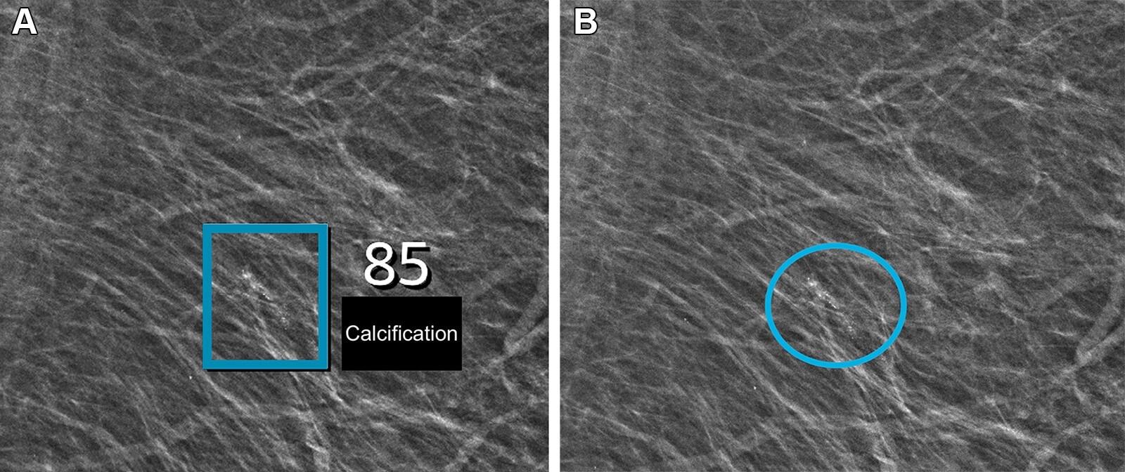
The AI system implemented to screen mammographs was trained using deep learning models to detect, highlight, and rate any suspicious calcifications or lesions observed in the mammogram. The AI tool then stratified the screenings across a score range of 1 to 10, indicating breast cancer probability.
A team of radiologists, consisting mainly of senior radiologists experienced in reading breast imaging results, read the mammograms for both cohorts. Before the implementation of the AI screening system, each screening was read by two radiologists, and the patient was recommended a clinical examination and needle biopsy only if both radiologists indicated the screening as warranting recall.
After the AI screening system was implemented, the mammograms that had a score lower than or equal to 5 were read by a senior radiologist who was aware that those mammograms underwent only one read. Those that warranted recall were then discussed with a second radiologist.
 Left mediolateral oblique full-field digital mammographic view in a 67-year-old woman with a Breast Imaging Reporting and Data System density of 1 who underwent screening with the artificial intelligence (AI) system. (A) Image shows AI-provided marking (square). The screening received a high AI examination score of 10, based on this area with arterial calcifications being given a score of 85 out of 100 by the AI system. (B) Same image as in A, but with findings by the radiologists. Because of the high AI examination score, the screening was double read by two radiologists, who determined that the arterial calcifications (circle) did not yield suspicions for breast cancer. The woman was not recalled for diagnostic assessment.
Results
The study found that implementing the AI-based screening system significantly lowered the workload for radiologists analyzing mammograms from a population-based breast cancer screening program while improving the screening performance.
The cohort that was screened before the implementation of the AI-based screening system consisted of over 60,000 women, while the cohort that was screened using the AI system had about 58,000 women. The AI screening resulted in an increase in breast cancer diagnoses (0.70% versus 0.82% before AI versus with AI, respectively) with a lower rate of false positives (2.39% versus 1.63%).
AI-based screening had a higher positive predictive value, and the percentage of invasive cancers was lower when AI-based methods were used for screening. Although the node-negative cancer percentage did not change, the other performance indicators showed that AI-based screening significantly improved performance. The reading workload was also found to have lowered by 33.5%.
Conclusions
To summarize, the study evaluated the effectiveness of an AI-based screening system in decreasing radiologists' workloads and improving screening performance in reading mammograms for biennial population-based breast cancer screening in Denmark.
The findings showed that the AI-based system significantly lowered the workload for radiologists while improving screening performance, supported by a substantial increase in breast cancer diagnoses and a significant decrease in false positive rates.
Journal reference:
- Lauritzen, A. D., Lillholm, M., Lynge, E., Nielsen, M., Karssemeijer, N., Vejborg, I., & Moy, L. (2024). Early indicators of the impact of using AI in mammography screening for breast cancer. Radiology, 311(3), e232479. DOI: 10.1148/radiol.232479, https://pubs.rsna.org/doi/10.1148/radiol.232479