Oxytocin is a hormone that plays a vital role in regulating social and emotional behaviors, cardiovascular functions, and energy intake. Various social and non-social conditions stimulate the endogenous oxytocin system, leading to increased neuropeptide synthesis in the hypothalamus and subsequent release within the brain and peripheral circulation.
Estimating oxytocin concentrations in peripheral samples, including blood, saliva, urine, and sweat, is generally preferred over cerebrospinal fluid puncture as the latter is an invasive method associated with higher health risks and intense medical care.
However, the sensitivity and reliability of measuring oxytocin concentration in saliva and urine samples remains uncertain. It is also unclear whether and under which conditions urinary oxytocin correlates with salivary oxytocin concentrations.
In this study, scientists have investigated whether urinary and salivary measurements of oxytocin and cortisol levels are sufficient to accurately capture physical exercise-induced fluctuations in their concentrations. They have included cortisol as a marker of hypothalamic-pituitary-adrenal (HPA) axis activation following exercise.
Study design
The study was conducted on 43 healthy adult male and female individuals who were divided into two groups.
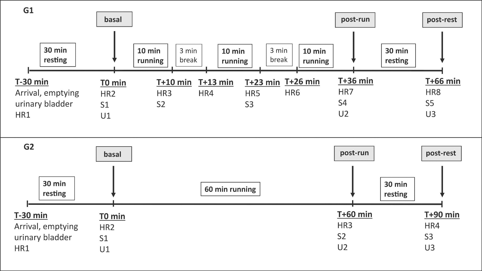
The first group (group 1) included 20 participants who were instructed to run three times for 10 minutes, with short intervals in between to provide heart rate and saliva samples. They also provided heart rate and urine and saliva samples at baseline and after running and resting periods.
The second group (group 2) included 23 experienced runners who were instructed to run continuously for 60 minutes at a fixed speed. They provided heart rate, urine samples, and saliva samples at baseline (pre-running), post-running, and 30-minute post-exercise resting periods.
Both urine and saliva samples were analyzed to measure oxytocin and cortisol concentrations. The effects of specific control predictors, including age, gender, cycle phase, and previous running experience of participants, were considered in the statistical analyses.
Important observations
The comparison of pre- and post-running heart rates revealed a significant induction in both study groups, with group 1 showing comparatively higher heart rates than group 2.
Running-induced changes in oxytocin concentration
Regarding urinary oxytocin concentration, a significant induction was observed at post-running and post-resting time points in both groups compared to that at pre-running time points. A significant reduction in oxytocin concentration was observed from post-running to post-resting time points. Overall, oxytocin concentrations were marginally higher in group 2 than in group 1.
Salivary oxytocin concentrations in both groups showed similar fluctuation patterns as urinary oxytocin concentrations. Oxytocin in saliva was elevated after just 10 minutes and peaked after 30 minutes of running.
Among control predictors, only age showed a significant negative association with urinary oxytocin concentrations but not with salivary oxytocin concentrations.
Running-induced changes in cortisol concentration
Urinary cortisol concentrations significantly increased from pre-running to post-resting time points in group 2. However, no significant differences in urinary cortisol concentrations were observed between pre-running, post-running, and post-resting time points in group 1.
No significant differences were observed between tested timepoints in both groups regarding salivary cortisol concentrations.
Among control predictors, previous running experience was found to influence urinary cortisol concentrations.
 Experimental timeline for group 1 (G1, upper panel) and group 2 (G2, lower panel). Pulse rate (HR), saliva samples (S) and urine samples (U) were taken as indicated.
Associations between urinary and salivary oxytocin and cortisol concentrations
Running-induced increase in urinary and salivary oxytocin concentration from pre-running to post-running timepoint showed significant positive associations in both study groups. However, no such association was observed for absolute oxytocin centration.
The running-induced increase in urinary and salivary cortisol concentration from the pre-running to the post-resting timepoint showed significant positive associations in both groups.
Regarding absolute cortisol concentrations, urine and saliva samples showed significant associations at the pre-running timepoint in group 1 and at the post-resting timepoint in group 2.
Study significance
The study shows that physical exercise can robustly stimulate oxytocin secretion in the periphery, as observed by significantly increased concentrations in both urine and saliva samples. Moreover, the study finds significant associations between post-exercise urinary and salivary oxytocin concentrations in both groups of runners, irrespective of running patterns and previous running experience.
These findings highlight that urine and saliva are adequate substitutes for blood sampling to monitor peripheral oxytocin concentrations, both during the oxytocin system's basal activity and in response to exercise.
The study findings also indicate that salivary sampling is associated with higher temporal resolution due to the provision of higher sampling frequency; however, urinary sampling is associated with higher signal strength and robustness.
Since differences in osmolarity can confound urinary hormone concentrations, the scientists advise correcting the measurement for creatinine concentration or specific gravity of the samples and excluding extreme outliers.