In this interview, News-Medical LifeSciences speaks with Professor Mark Lewis, Co-Founder and CEO of Myomaker Bio, about the development of lab-grown human muscle platforms for preclinical drug testing.
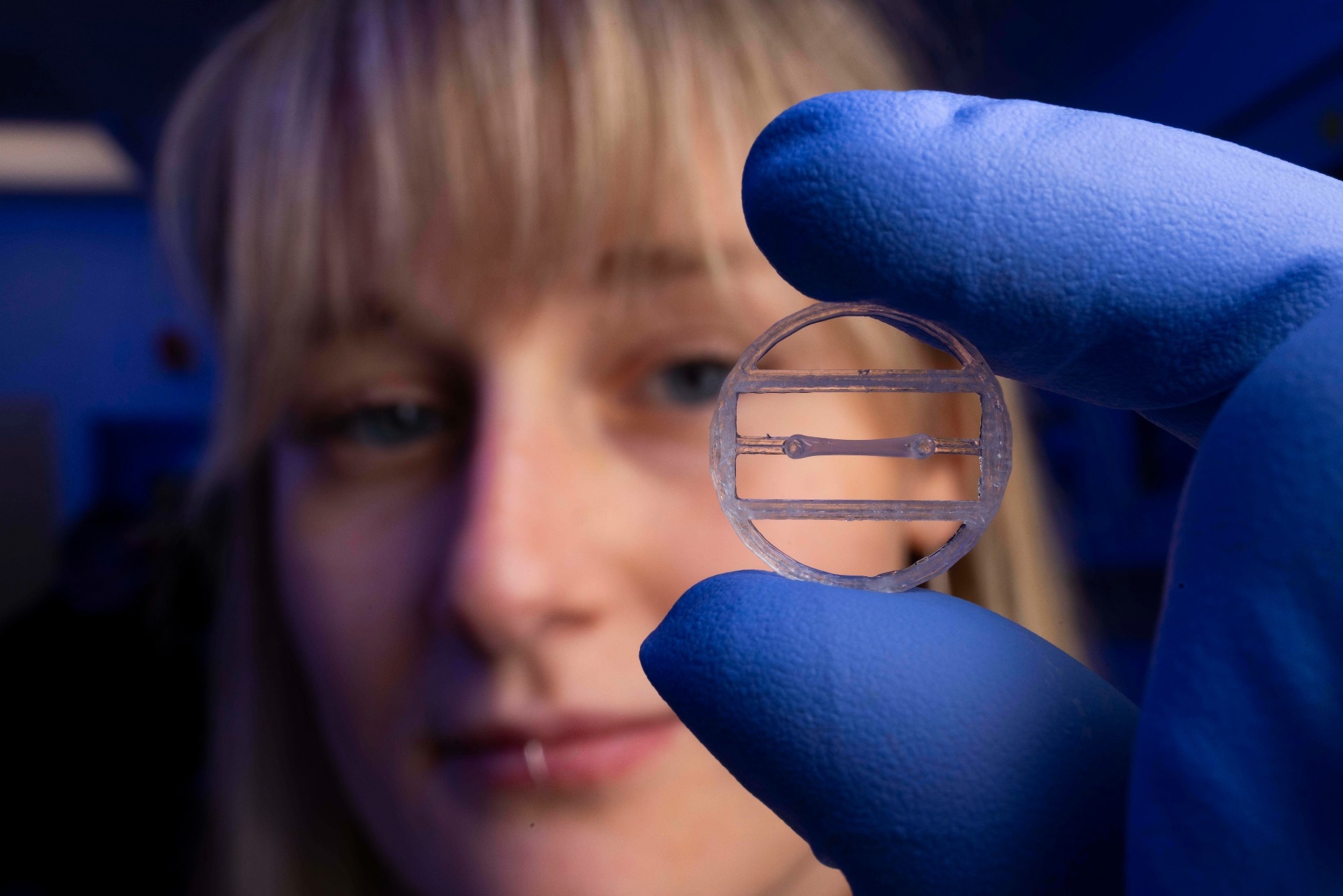 Image credit: Myomaker Bio
Can you explain what Myomaker Bio's lab-grown human muscle platform is and the challenge it aims to address in drug development?
Myomaker Bio develops three-dimensional human-derived muscle organs and complex tissue systems that can be incorporated into bespoke preclinical testing assays. These models allow pharmaceutical and biotechnology companies to study how candidate drugs interact with human muscle tissue under biologically relevant conditions.
The core challenge we are addressing is the limited predictive value of many traditional preclinical models, particularly animal studies. Human-relevant tissue systems can provide earlier insight into drug safety, efficacy, and underlying mechanisms, helping to reduce risk and accelerate development timelines.
How did your research at Loughborough University evolve into the technology that now underpins Myomaker Bio?
I’ve been working in this area for around 20 years; it’s essentially been my life’s work. Over that time, my team and I have developed extensive expertise in creating novel muscle models, and for at least the past decade, we’ve been exploring how to commercialize that work and scale its impact.
Together with my co-founder, Dr. Andrew Capel, we felt the timing was right to establish Myomaker Bio. Both personally and technologically, we had reached the point where spinning out of Loughborough University and seeking investment were the natural next steps in advancing the platform.
When you say your engineered tissues replicate the structure and function of real skeletal muscle, what does that mean in practical terms?
Our lab-grown muscle closely resembles real skeletal muscle both structurally and functionally. It not only looks like skeletal muscle tissue under analysis, but also behaves in the same way biologically.
We’ve demonstrated this using a range of anatomical and functional assessment techniques, showing that the tissues develop the same organized structure and contractile behavior seen in human skeletal muscle.
Could you walk us through how these lab-grown muscle tissues are created?
We typically begin with primary human muscle biopsies taken from the vastus lateralis, one of the quadriceps muscles. The tissue is minced and placed into a culture flask. The cells that migrate out from the tissue are muscle stem cells, known as satellite cells, whose natural role is to repair damaged muscle and support muscle growth by restoring or adding nuclei to muscle fibers.
Once isolated, these cells are expanded for use within our system. In the laboratory, we effectively recapitulate the natural muscle regeneration process by activating satellite cells and guiding their development into mature muscle tissue.
The cells are then suspended in a collagen-Matrigel matrix and placed into small, circular, 3D-printed frames with two anchor points. As the matrix solidifies into a gel, the tissue aligns between the posts and begins to contract. Over time, this produces multinucleated myotubes and mature muscle fibers, which are surrounded by collagen and anchored between two fixed points, creating organized, functional muscle tissue.
How closely do these models reflect real human muscle responses to exercise, injury, regeneration, and pharmacological treatments?
Our muscle models behave in ways that closely reflect normal human muscle biology, and we’ve established multiple validation points to demonstrate this.
We have validated them using compounds such as leucine, insulin, testosterone, and Resolvin E1, demonstrating predictable changes in muscle morphology and function. The tissues can also be mechanically or electrically stimulated to simulate exercise, thereby altering force production and muscle characteristics.
We have developed disease models of metabolic disorders by exposing tissues to excess fatty acids, creating metabolic dysfunction similar to that observed under high-fat diet conditions. We can also induce injury using compounds such as barium chloride and observe subsequent regeneration, enabling detailed investigation of repair mechanisms in a highly controlled environment.
What have you learned about how your human muscle platform compares with traditional animal models?
Drug development remains slow, expensive, and heavily reliant on animal models that do not always predict human outcomes accurately. Our human muscle platforms are designed to help bridge that gap by providing more human-relevant data earlier in the development process.
By improving the predictive power of preclinical testing, our models have the potential to make drug development faster, more efficient, and ultimately more successful. Our ambition is for them to significantly shorten development timelines and increase the likelihood that new therapies will succeed in the clinic.
In studies conducted with industry partners, compounds known to induce muscle toxicity in animal models have also produced comparable toxic effects in our human-derived systems. This provides confidence that the platform can identify clinically relevant responses while offering a more direct representation of human tissue biology. Ultimately, we believe human tissues should be used to predict human outcomes.
How could these platforms improve decision-making for pharmaceutical companies developing muscle-targeting therapies?
Muscle is one of the most commonly affected organs during preclinical safety studies. Our platform allows researchers to assess muscle-specific toxicity directly in human tissue rather than relying solely on indirect observations from animal models.
The ability to generate data rapidly means researchers can evaluate compounds earlier, optimize lead candidates more efficiently, and make informed development decisions based on human-relevant evidence. This can reduce risk, improve efficiency, and potentially shorten development timelines.
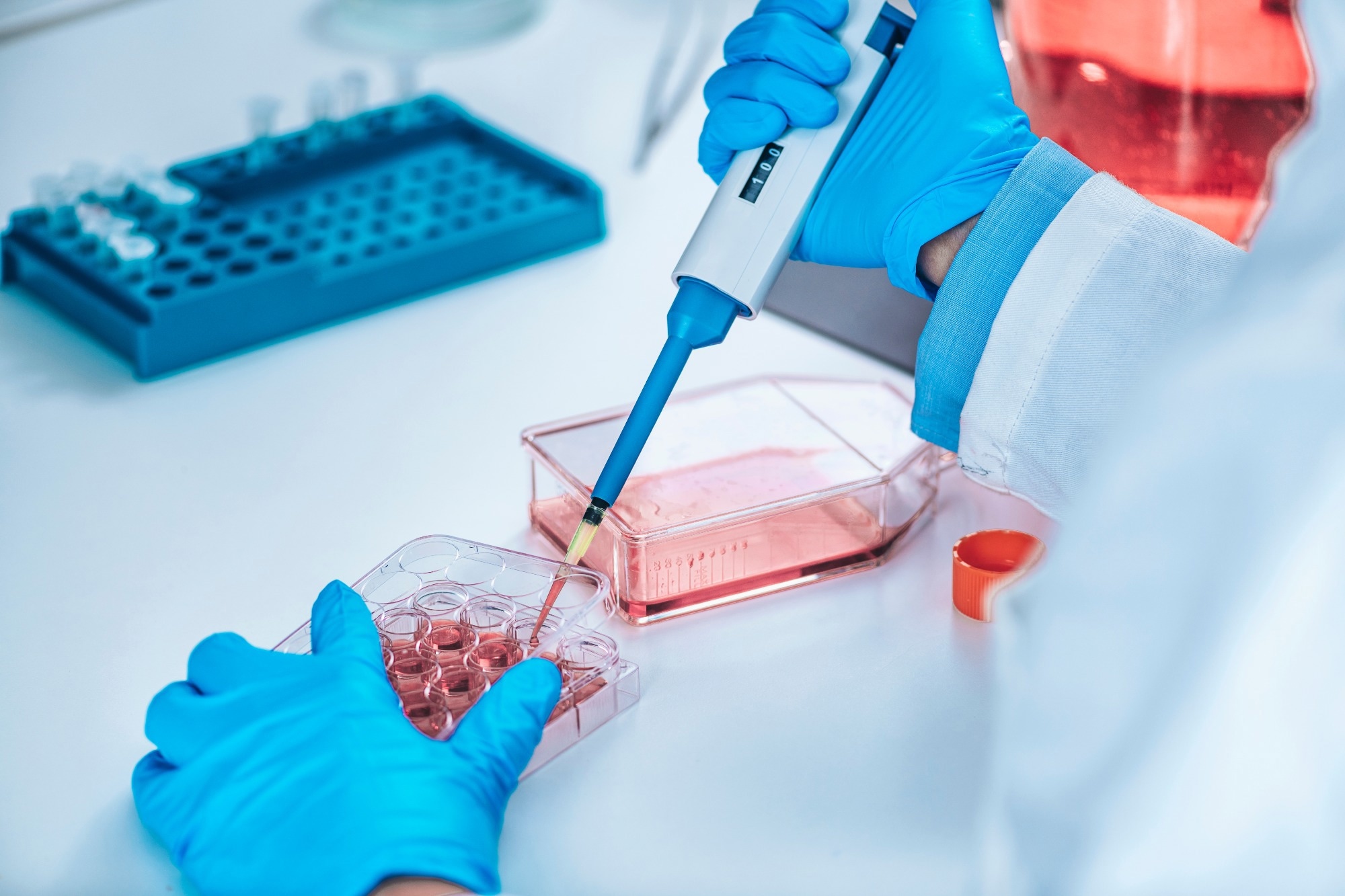 Human-derived muscle tissues can be used to evaluate drug safety, efficacy, and muscle-specific toxicity in a controlled laboratory setting, providing pharmaceutical companies with earlier, more clinically relevant data to support preclinical decision-making. Image credit: Microgen/Shutterstock.com
What were the biggest challenges in transforming this technology from an academic research platform into a commercial product?
One of the biggest initial challenges was making the product faster to produce, more cost-effective, and smaller in scale, while also ensuring it could be reproduced consistently and reliably. Academic systems are often designed for discovery, whereas commercial platforms must be standardized and scalable.
Fortunately, much of this work had already been completed during the research and funding stages prior to Myomaker Bio's founding. That early groundwork has been critical in helping us transition from an academic research platform into something that can be manufactured and deployed at a commercial scale.
Following your recent investment, what are the next steps to scale the platform and expand its capabilities?
From an operational perspective, the immediate challenge is to scale production while maintaining consistent, reproducible outputs across multiple client programs.
The next challenge will be expanding the range of assays and disease models we can support. For example, can we build models using muscle samples from patients in different regions of the world, including those with muscular dystrophy? I believe we can.
Beyond that, the greater challenge is modeling more complex conditions such as motor neuron disease, where multiple biological systems interact. Recreating those disease environments in a meaningful and physiologically relevant way will be essential to deepening our understanding of those conditions.
Where can readers find more information?
About the researcher
Professor Mark Lewis is Co-Founder and CEO of Myomaker Bio. He has held leadership positions at several academic institutions across different spheres of higher education while maintaining his status as a fully research-active academic. He has been involved in line, change, and group management and also held a number of directorships and consultancies with companies in the life sciences sector, including involvement in multiple projects in a leadership capacity within these roles.
Alongside this, he has dedicated more than 25 years to the development and applications of human muscle organs. He has authored and co-authored over 50 publications in the skeletal muscle field
Journal references:
- Fleming, J.W., Capel, A.J., Rimington, R.P., Player, D.J., Stolzing, A. & Lewis, M.P., (2019). Functional regeneration of tissue engineered skeletal muscle in vitro is dependent on the inclusion of basement membrane proteins. Cytoskeleton, 76(6), pp.371–382. DOI: https://doi.org/10.1002/cm.21553
- Martin, N.R.W., Turner, M.C., Farrington, R., Player, D.J. & Lewis, M.P., (2017). Leucine elicits myotube hypertrophy and enhances maximal contractile force in tissue engineered skeletal muscle in vitro. Journal of Cellular Physiology, 232(10), pp.2788–2797. DOI: https://doi.org/10.1002/jcp.25960
- Rimington, R.P., Fleming, J.W., Capel, A.J., Wheeler, P.C. & Lewis, M.P., (2021). Bioengineered model of the human motor unit with physiologically functional neuromuscular junctions. Scientific Reports, 11(1), 11695. DOI: https://doi.org/10.1038/s41598-021-91203-5
- Turner, M.C., Martin, N.R.W., Player, D.J., Ferguson, R.A., Wheeler, P., Green, C.J., Akam, E.C. & Lewis, M.P., (2020). Characterising hyperinsulinaemia-induced insulin resistance in human skeletal muscle cells. Journal of Molecular Endocrinology, 64(3), pp.125–132. DOI: https://doi.org/10.1530/JME-19-0169
- Baker, L.A., Martin, N.R.W., Kimber, M.C., Pritchard, G.J., Lindley, M.R. & Lewis, M.P., (2018). Resolvin E1 attenuates LPS-induced inflammation and subsequent atrophy in C2C12 myotubes. Journal of Cellular Biochemistry, 119(7), pp.6094–6103. DOI: https://doi.org/10.1002/jcb.26807
- Hughes, D.C., Stewart, C.E., Sculthorpe, N., Dugdale, H.F., Yousefian, F., Lewis, M.P. & Sharples, A.P., (2015). Testosterone enables growth and hypertrophy in fusion impaired myoblasts that display myotube atrophy: deciphering the role of androgen and IGF-I receptors. Biogerontology, 17(3), pp.619–639. DOI: https://doi.org/10.1007/s10522-015-9621-9