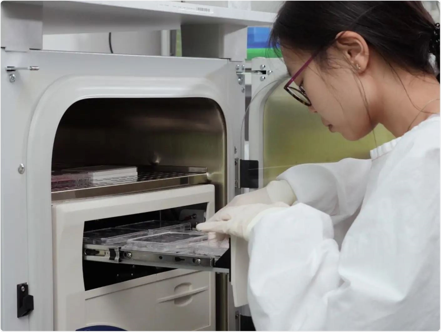
 Citation: Zhang, X., Morten, B.C., Scott, R.J., Avery-Kiejda, K.A. A Simple Migration/Invasion Workflow Using an Automated Live-cell Imager. J. Vis. Exp. (144), e59042, doi:10.3791/59042 (2019).
Cell migration and invasion processes play a role in many normal and pathological cellular processes. One of these pathological processes is tumor metastasis. In vitro assays are an excellent way to study both 2D and 3D cell movements to help you better understand cancer cell metastasis.
Researchers at Hunter Medical Research Institute and Priority Research Centre for Cancer Research have developed a simultaneous migration and invasion assay protocol that offers you a time-efficient, simple, and reproducible experimental option to study cell migration and invasion, and thus cancer cell metastasis.
Journal abstract: Cancer cell mobility is crucial for the initiation of metastasis. Therefore, investigation of the cell movement and invasive capacity is of great significance. Migration assays provide basic insight of cell movement at a 2D level, whereas invasion assays are more physiologically relevant, mimicking in vivo cancer cell dislodgment from the original site and invading through the extracellular matrix. The current protocol provides a single workflow for migration and invasion assays. Together with the integrated automated microscopic camera for real-time HD images and built-in analysis module, it gives researchers a time-efficient, simple, and reproducible experimental option. This protocol also includes substitutions for the consumables and alternative analysis methods for users to choose from.