The medical industry is a new frontier in Raman spectroscopy. Raman has been employed in dental and cancer studies in recent years, and its success is being expanded into Point-of-Care (POC) applications.
This article provides a summary of some new and intriguing research into using Raman spectroscopy to detect malignant tissue, disease biomarkers, and disease-causing pathogens.
Detecting bone infection with a handheld Raman spectrometer
Complication from infection is a serious concern when using human bone grafts in musculoskeletal surgery. Bone-related infections are typically caused by Staphylococcus epidermidis and Staphylococcus aureus, which are challenging to treat effectively.
Image Credit: Adobe Stock
Detecting staph bacteria on graft material and distinguishing between healthy and diseased bone are both critical in preventing infection.
Laboratory culture typically takes 7–10 days to produce results and is susceptible to contamination during transportation and testing. In positive cases, the patient must be treated retroactively with large amounts of antibiotics.
On-site analysis is an ideal solution to this problem since it allows the surgical team to detect and avoid diseased bone on the spot.
A study group in Austria recently demonstrated a successful distinction between healthy and infected bone samples, as well as between two strains of staph bacteria, utilizing a handheld MIRA Raman spectrometer.1
Their method analyzed the fingerprint Raman bands of phosphates, amides, and collagens, as well as their varying intensity and peak width ratios, to differentiate between healthy and diseased bone. Principal component analysis (PCA) facilitated optical analysis and was used to distinguish between staph strains more precisely.
The Austrian group appreciated MIRA’s lightweight, small, and battery-powered design and the fact that Raman spectroscopy requires minimum sample preparation and produces rapid results. Testing only requires a small bone sample for in-situ testing during surgery and offers quick and precise results directly in the operating room.
Raman spectroscopy for cancer detection
Raman’s molecular fingerprint spectra are sensitive enough to identify the chemical changes associated with disease. Thus, compact Raman spectrometers can help surgeons examine tumors during surgical procedures, allowing for rapid decision-making.
Application examples for breast cancer and pancreatic cancer are presented in the sections below.

Image Credit: Adobe Stock
Traditional Raman spectroscopy in breast cancer assessment
When breast cancer is suspected, one surgery is usually needed for a biopsy and another for the removal of cancerous tumors. The capability to analyze suspected tissues during preliminary surgery allows for immediate removal if necessary.
The benefit to the patient and the medical sector is immeasurable—and research indicates that Raman spectroscopy may be able to meet this demand for some types of cancer.
Raman spectroscopy is sensitive enough to detect tissue changes caused by various types of cancer. For instance, there are very subtle differences in Raman spectra from healthy breast tissue and malignant tumors.
Researchers in the United Kingdom employed a high-resolution i-Raman laboratory device (Figure 1) and multivariate methods, including PCA, to successfully distinguish between healthy and cancerous tissues.2
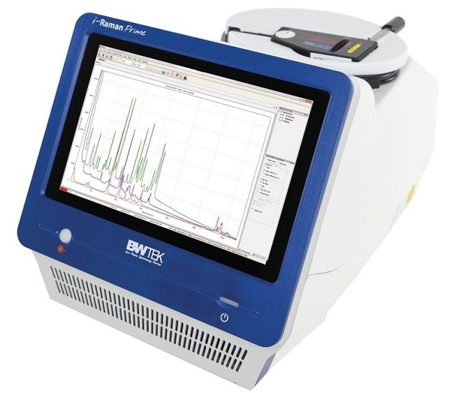
Figure 1. The i-Raman Prime 785S Portable Raman Spectrometer from Metrohm. Image Credit: Metrohm Middle East FZC
SERS for detection and measurement of pancreatic cancer biomarkers
SERS (surface-enhanced Raman spectroscopy) can be the best option when Raman is not suitable for analysis. This can be the case in situations where the target exists in a complex sample matrix or due to the fluorescence of carbon-based molecules. SERS improves the Raman signal but not the competing fluorescence signal.
The SERS effect allows for sensitive detection of analytes at mg/L levels—sometimes as low as µg/L. Finally, SERS peaks are sharp and well-defined, enabling effective detection and identification of target analytes.
Pancreatic cancer is fatal, partly because it is difficult to diagnose. However, several biomarkers are found at increased levels in about 75 % of positive cases.3 These can be detected using enzyme-linked immunosorbent assays (ELISA), which test a range of biomarkers such as antibodies, antigens, and proteins.
In an emerging technique, an i-Raman laboratory spectrometer was used at the University of Utah for SERS analysis in combination with ELISA to identify an antigen linked to pancreatic cancer.2
The SERS signal was produced by a reporter molecule that was complexed with both a gold nanoparticle and the target analyte in an otherwise standard lateral-flow or sandwich immunoassay (Figure 2). This extremely accurate technique allows for the sensitive detection and even quantification of the biomarker of interest.
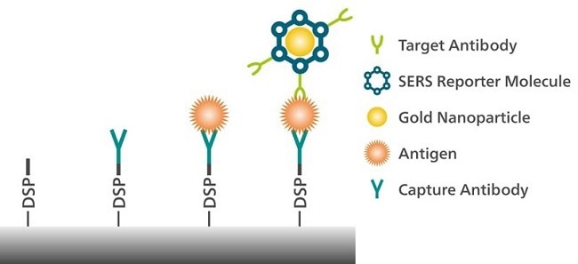
Figure 2. Detection of an antigen associated with pancreatic cancer is possible with surface-enhanced Raman spectroscopy (SERS). Image Credit: Metrohm Middle East FZC
Rapid, point-of-care (POC) assay for femtogram-level detection of COVID-19
Researchers from the University of Wyoming used an alternative ELISA technique to detect antigen biomarkers linked with COVID-19 infection.4 This study used a magnetic nanoparticle-supported assay to concentrate the target biomarker in solution for subsequent SERS detection with MIRA XTR (Figure 3).
It outperformed commercial lateral flow assays in terms of sensitivity, compatibility with both solvent and saliva samples, adaptability to new viral variants, and highly sensitive point-of-care COVID-19 diagnosis.
Lateral flow immunoassays produce reasonably fast results. However, they only deliver nanogram-level detection and have quantification restrictions. In contrast, the SERS-based ELISA is sensitive to femtogram quantities of antigen and provides rapid results at the point-of-care using a commercial handheld Raman instrument.
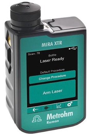
Figure 3. MIRA XTR, a handheld Raman spectrometer from Metrohm. Image Credit: Metrohm Middle East FZC
Multiplex immunophenotyping of blood and breast cancer cells with Raman spectroscopy
Another study used MIRA DS to test a portable SERS-based ELISA for immunophenotyping various types of red blood and breast cancer cell surfaces.5 Distinguishing between healthy and diseased cells, as well as detecting several biotargets in a single sample, can help with the informed treatment of several types of breast cancer.
Image Credit: Adobe Stock
This assay demonstrated higher specificity, sensitivity, and repeatability for immunophenotyping in diverse cell types using a smaller analysis sample volume than traditional multiplex immunoassays. It was also less labor-intensive and technically easier to perform.
The authors praised MIRA’s Orbital Raster Scan for enhancing the sensitivity of their assay by interrogating a larger area and taking spatially averaged measurements.
Conventional multiplexed flow tests may be limited by the availability of different colored stains and the interpretation of results. They are also associated with a large laboratory footprint.
In comparison, this approach based on handheld Raman spectroscopy has the potential to produce accurate POC results with short sample-to-result time, multiplexing capabilities, and very compact equipment.
Easy detection of enzymes with the electrochemical-SERS effect
A very different SERS method for characterizing biological molecules has been reported by Metrohm.6
Electrochemical SERS (EC-SERS) allows for two experiments at once: electrochemical activation of SERS features of silver electrodes (Ag SPEs) followed by spectroscopic detection of the sample (with SPELECRAMAN638, Figure 4).
The SERS substrate is produced in situ from silver electrodes (both screen-printed and conventional).
This is performed in the presence of the analyte while being continuously interrogated with Raman to optimize the detection of SERS-active species. The 638 nm excitation produces a strong SERS effect while minimizing the risk of sample damage and fluorescence.
The structure of enzymes (and their role in disease), like aldehyde dehydrogenase (ALDH), can help when it comes to understanding diseases.
Using EC-SERS, application scientists identified previously unreported fingerprint Raman bands of ALDH in solution. Similarly, the redox states of cytochrome C reveal information regarding electron transport across cell membranes.7
During the EC experiment, cytochrome C changes oxidation and conformation states, and these redox states have distinguishable SERS spectra.
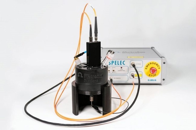
Figure 4. The SPELECRAMAN638 instrument from Metrohm performs spectroelectrochemical Raman measurements using a 638 nm laser. Image Credit: Metrohm Middle East FZC
Conclusion
Research teams around the world are harnessing Raman technology in innovative ways. Leveraging its benefits, such as trace detection, high sensitivity, compact size, and rapid results, this technology is making significant strides in identifying cancerous tissues, detecting disease biomarkers, and pinpointing pathogens that cause illnesses. The outcomes of these applications are both exciting and promising.
References
- Lindtner, R. A.; Wurm, A.; Pirchner, E.; et al. Enhancing Bone Infection Diagnosis with Raman Handheld Spectroscopy: Pathogen Discrimination and Diagnostic Potential. IJMS 2023, 25 (1), 541. DOI:10.3390/ijms25010541
- Thomas, R.; Bakeev, K.; Claybourn, M.; Chimenti, R. The Use of Raman Spectroscopy in Cancer Diagnostics. Spectroscopy 2013, 28 (9), 36–43.
- Goonetilleke, K. S.; Siriwardena, A. K. Systematic Review of Carbohydrate Antigen (CA 19-9) as a Biochemical Marker in the Diagnosis of Pancreatic Cancer. Eur J Surg Oncol 2007, 33 (3), 266–270. DOI:10.1016/j.ejso.2006.10.004
- Antoine, D.; Mohammadi, M.; Vitt, M.; et al. Rapid, Point-of-Care ScFv-SERS Assay for Femtogram Level Detection of SARS-CoV-2. ACS Sens. 2022, 7 (3), 866–873. DOI:10.1021/acssensors.1c02664
- Wang, J.; Koo, K. M.; Trau, M. Tetraplex Immunophenotyping of Cell Surface Proteomes via Synthesized Plasmonic Nanotags and Portable Raman Spectroscopy. Anal. Chem. 2022, 94 (43), 14906–14916. DOI:10.1021/acs.analchem.2c02262
- Metrohm AG. Easy Detection of Enzymes with the Electrochemical-SERS Effect; AN-RA-008; Metrohm AG: Herisau, Switzerland, 2023.
- Brazhe, N. A.; Evlyukhin, A. B.; Goodilin, E. A.; et al. Probing Cytochrome c in Living Mitochondria with Surface-Enhanced Raman Spectroscopy. Sci Rep 2015, 5 (1), 13793. DOI:10.1038/srep13793
About Metrohm Middle East FZC
Metrohm Middle East FZC is a subsidiary of Metrohm AG, Switzerland, located at Sharjah Airport International Free Zone, UAE. Metrohm’s Product range involves both Lab instruments & online Analysers for various industry segments - Water, Petrochemical, Pharmaceutical, Food and Beverages, Environmental Analysis, Chemical analysis, Research and development, Educational Institutions, Sewage Treatment and more.
MME has a team of well trained; knowledgeable and experienced team specialized in Product Management, Service and Application support. Apart from extending our support to distributors, MME also provides direct support and training to our customers on site or at our world class Regional Support Centre.
Sponsored Content Policy: News-Medical.net publishes articles and related content that may be derived from sources where we have existing commercial relationships, provided such content adds value to the core editorial ethos of News-Medical.Net which is to educate and inform site visitors interested in medical research, science, medical devices and treatments.