Diplonemid flagellates are among the most abundant marine microeukaryote species in world ocean habitats.1 Diplonemids are recognized for their complex organelle structure and mitochondrial DNA.2
Although abundant in the ocean, mechanistic understanding is ‘fragmentary,’ with most research focused on organelle structure and being negligent of a cell-level approach. In this article, you will read about how researchers built a detailed 3D ultrastructural characterization of the entire diploid cell, particularly about the mitochondria structure and the discovery of a new organelle.
Image Credit: Shutterstock AI Generator
Looking beyond the cell
Understanding the cell biology of a eukaryotic group is crucial for accurately interpreting ecological and molecular information. Despite diplonemid protists being among the most species-diverse lineages of marine eukaryotes, only a limited and incomplete amount of data is available regarding the cellular structure of this varied group.
Researchers from the Czech Academy of Sciences and the University of South Bohemia (Czech Republic) have isolated a new diplonemid strain called Lacrimia vacuolate sp. nov, derived from Japan's sea.3
They used serial block-face scanning electron microscopy (SBF-SEM) technology, array tomography, and fluorescence microscopy combined with image analysis software to obtain the first three-dimensional reconstruction of a diplonemid cell.
This innovative workflow enabled researchers to identify a unique mitochondrial morphology and a rich endomembrane system composed of various vesicles and vacuoles. Notably, the discovery includes a new organelle named colv (Center for Organization of Layered Vesicles).
Unique mitochondrial morphology identified
A 3D reconstruction of five cells revealed that each cell contains a single unbranched mitochondrion. This mitochondrion is largely situated just beneath the microtubular corset, except for a small branch that connects with the cytopharynx (Ph).
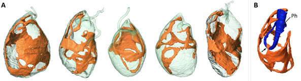
Fig.1: Shape and detailed structure of the mitochondrion. A) Surface rendering of five cells reconstructed from SBF-SEM data, each showing a single interconnected mitochondrion (orange). B) Connection point between the mitochondrion and the cytopharynx (Ph). Image Credit: doi: 10.1128/mbio.01921-23.
Although the mitochondrion covers a large area, it often narrows down to thin sections with closely juxtaposed membranes lacking visible cristae or matrix. These thin sections link thicker regions that contain dense matrix and prominent DNA aggregates, as well as cristae structures. The cristae come in various shapes, including long parallel layers, short straight or curved layers, and blob-like forms.
Novel organelle discovered
One of the most striking findings in the ultrastructural characterization of Lacrimia vacuolate is the discovery of a novel endomembrane organelle named the ‘Center for Organization of Layered Vesicles’ (colv).
Analysis of ultrathin TEM sections showed this organelle is in the front region of the cell, adjacent to the cytopharynx (Fig. 2). The organelle is constituted of both short and long flat vesicles.
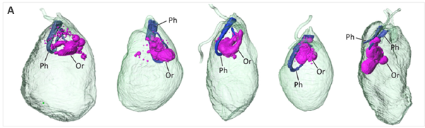
Fig.2: 3D reconstruction of 5 cells which shows colv organelle and the cytopharynx. Image Credit: doi: 10.1128/mbio.01921-23.
Several simple, small vesicles, as well as larger double-membrane ring-shaped vesicles, were also linked to the organelle. Live cells were incubated with lanthanoid nanocrystals to verify the connection between the organelle and vesicles derived from the cytopharynx. The cells were then prepared for TEM analysis.
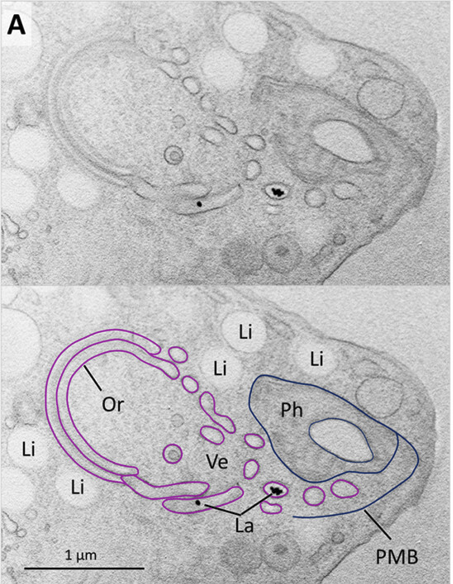

Fig.3: The interaction between colv organelles and cellular structures. A) TEM images of cells treated with lanthanoid nanocrystals (La). B) 3D reconstruction of 2 μm-thick slice from array tomography data set shows different organelles vacuoles and vesicles. Nucleus (Nu), mitochondrion (Mt), lipid droplets (Li), posterior vacuole(PV), cytopharynx (Ph) Colv organelle (Or) and smaller vacuole (Va). Image Credit: doi: 10.1128/mbio.01921-23.
The nanocrystals were clearly visible within the pharyngeal lumen and cytopharynx-derived vesicles, including the ring-shaped vesicles, where the crystals were observed inside the lumen.
Characterizing the Lacrimia vacuolate structure showcases the excellent potential for ultrastructural and 3D reconstruction. These findings highlight how high-resolution imaging can characterize function and identify emergent properties, mechanisms, and even novel organelles. The possibilities for this technology move beyond marine life, as it is possible to apply it to the visualization of any cell or organelle type, which enables better derivation of structure-function relationships.
Thermo Fisher Scientific technology
In this study, the Thermo Scientific™ Amira™ software was utilized to process the TEM and SBF-SEM data, allowing the creation of 3D ultrastructure. Amira enables the reconstruction of high-quality images gained by electron microscopy technologies as well as other technologies such as magnetic resonance imaging (MRI), X-ray, and optical microscopy for 3D modeling.
Amira Software allows researchers to enhance efficiency and save time by managing various stages, including visualization, segmentation, and intricate morphological measurements.
Amira for Cell biology webinar teaser
Watch on-demand how to maximize your cell biology images with Amira Software
References and Further Reading
- [1 ] Flegontova O, Flegontov P, Malviya S, Audic S, Wincker P, de Vargas C, et al. Extreme diversity of diplonemid eukaryotes in the ocean. Curr Biol. 2016;26(22):3060–5.
- Marande W. et al. (2005) Unique mitochondrial genome structure in diplonemids, the sister group of kinetoplastids. Eukaryot Cell. doi: 10.1128/EC.4.6.1137-1146.2005.
- Tashyreva D. et al. (2023) Ultrastructure and 3D reconstruction of a diplonemid protist (Diplonemea) and its novel membranous organelle. mBio doi: 10.1128/mbio.01921-23.
- Tashyreva D, et al. (2022.) “Diplonemids – a review on “new” flagellates on the oceanic block” Protist. doi;173:125868.
- Amira Software for cell biology | Thermo Fisher Scientific - DE

Sponsored Content Policy: News-Medical.net publishes articles and related content that may be derived from sources where we have existing commercial relationships, provided such content adds value to the core editorial ethos of News-Medical.Net which is to educate and inform site visitors interested in medical research, science, medical devices and treatments.