Cryopreservation temporarily disrupts living cell systems. While membrane-integrity or dye-exclusion assays provide a snapshot of live cell counts at a given time post-thaw, they do not show whether the culture can attach, grow, retain its identity, or perform the activity needed for the experiment.
That gap is even more crucial in induced pluripotent stem cell (iPSC) and three-dimensional cell culture workflows. Although it contains viable cells, a culture may show poor attachment, slow expansion, lineage marker loss, altered organization, or failure to deliver the anticipated electrical, secretory, or contractile response.
Post-thaw recovery requires assessing cell survival and the model’s ability to perform as needed after recovery. This can be addressed systematically in four phases: survival, culture re-establishment, model integrity, and function.
A four-stage view of post-thaw recovery
Post-thaw recovery is a process rather than a single measurement. Four stages guide the culture from thawing to experimental application: verifying survival, checking culture re-establishment, confirming model identity and organization, and assessing the specific function needed for the intended application. The questions, testing times, and readouts presented serve as example recommendations and should be modified based on the specific model and target application.
Source: NIPPON Genetics EUROPE GmbH
| 1. Survival |
2. Culture re-establishment |
3. Model integrity |
4. Function |
Confirm cell survival
When: immediately after thawing |
Check culture recovery
When: during early post-thaw culture |
Verify identity and organization
When: after the culture has re-established |
Test the required activity
When: at the intended point of use |
Example readouts
Viability
Viable cell yield
Membrane integrity |
Example readouts
Attachment or aggregate reformation
Morphology and outgrowth
Return to growth |
Example readouts
Lineage or phenotype markers
Cell composition or histology
3D organization |
Example readouts
Secretion or uptake
Electrical or contractile activity
Response to a defined stimulus |
This table is a versatile planning guide, not a fixed test panel. For each relevant stage, select assessment timing and readouts based on the model, and define acceptance standards to determine whether the culture is ready to proceed to the next phase or its intended experimental application.
From surviving the thaw to working as intended
The evaluation thus extends beyond an initial live-cell count. After thawing, cultures from various cell models were assessed for their ability to re-establish growth, maintain model-specific properties, and recover the functional characteristics needed for their intended use.
The following studies evaluated these recovery parameters under model-specific freezing conditions, recovery periods, and experimental endpoints. Together, they show how Bambanker™ supported workflows that address the four phases of post-thaw recovery: survival, culture re-establishment, model integrity, and function.
iPSC-derived dopaminergic neurospheres
In a study by Hiramatsu et al., day-28 iPSC-derived dopaminergic progenitor neurospheres were cryopreserved using Bambanker™ hRM with a Proton Freezer protocol.1 Following thawing, the scientists evaluated viability, neurite extension, dopaminergic lineage markers, electrical activity, dopamine release, and performance after transplantation in a 6-hydroxydopamine-lesioned rat model.
Following a seven-day recovery period, fresh Day-35 neurospheres and neurospheres frozen on Day 28 displayed a comparably dense, rounded architecture, with broad staining for the floor-plate and dopaminergic lineage markers FOXA2, NURR1, and TH (Figure 1a).
The quantitative plots reveal closely overlapping marker-positive fractions in fresh and cryopreserved cultures, with no substantial variations across the four replicates (Figure 1b). Collectively, these images and counts indicate that the post-thaw spheres preserved their targeted dopaminergic identity rather than merely containing viable cells.
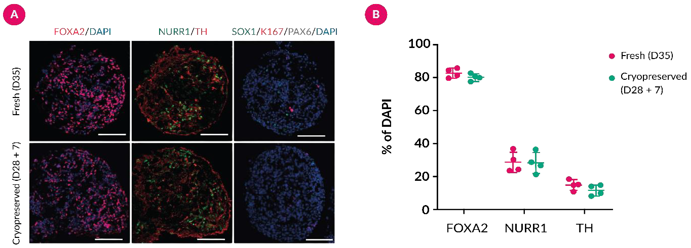
Figure 1. Dopaminergic lineage identity is maintained after cryopreservation. (A) Representative immunofluorescence images of fresh Day-35 neurospheres and neurospheres cryopreserved on Day 28 and recovered for seven days. Both conditions show widespread FOXA2 and NURR1 staining and TH-positive neuronal areas around DAPI-labelled nuclei, with no obvious loss of overall sphere organization after thawing. Scale bars: 100 μm. (B) The proportions of FOXA2-, NURR1- and TH-positive cells overlap between fresh and cryopreserved groups (n = 4; mean ± SD), and no significant differences were detected. Image Credit: NIPPON Genetics EUROPE GmbH
Multielectrode-array data from functional testing show similar spike amplitudes in fresh and cryopreserved neurospheres, signifying that the recovered neuronal networks remained electrically active.
The dopamine-release plot similarly demonstrates a comparable increase following high-potassium stimulation when normalized to DNA content. Neither endpoint exhibited significant differences between groups, supporting preservation of both excitability and stimulus-dependent dopaminergic secretion post-thaw (Figure 2).
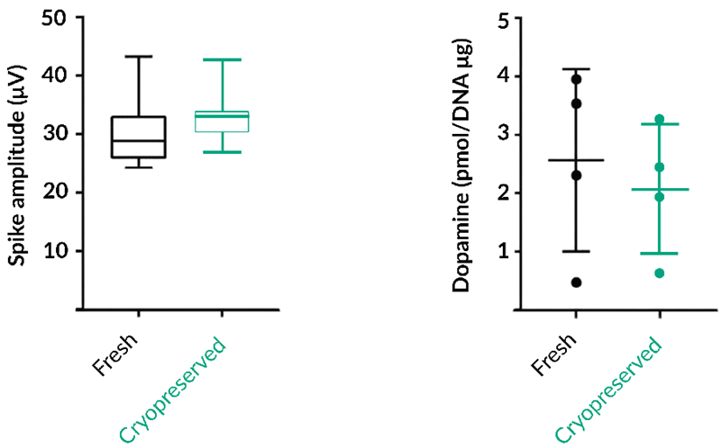
Figure 2. Post-thaw neurospheres retain in vitro neuronal function. The left panel compares spike amplitude measured by multielectrode array on Day 42; the distributions for fresh and cryopreserved neurospheres are similar, showing preserved electrical activity after recovery. The right panel shows high-potassium-induced dopamine release on Day 56, normalized to DNA content. Fresh and cryopreserved cultures display comparable stimulated release (n = 4; mean ± SD), with no significant difference between groups. Image Credit: NIPPON Genetics EUROPE GmbH
After confirming retention of neuronal activity and dopamine release in vitro, the scientists next assessed whether the recovered cells also maintained function after transplantation.
Vehicle-treated 6-OHDA-lesioned rats continued to show high levels of rotational behavior, while the curves for animals receiving fresh neurospheres or either dose of cryopreserved neurospheres declined over time. By Week 20, methamphetamine-induced rotations were significantly lower in all three cell-treated groups compared to the vehicle group (Figure 3).
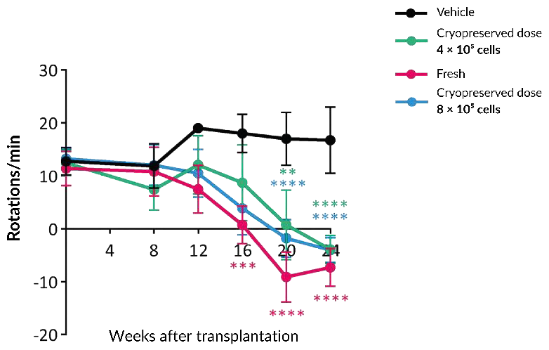
Figure 3. Cryopreserved neurospheres improve abnormal rotational behavior after transplantation. Methamphetamine-induced rotations were followed over 20 weeks in 6-OHDA-lesioned rats receiving vehicle, fresh neurospheres (4 × 10⁵ cells) or cryopreserved neurospheres (4 × 10⁵ or 8 × 10⁵ cells). Vehicle-treated animals maintain the highest rotation counts, while all cell-treated groups show a progressive reduction and separate from vehicle by Week 20 (n = 4–7; mean ± SD). Two-way ANOVA with Tukey’s test: **P < 0.01, ***P < 0.001, ****P < 0.0001. Image Credit: NIPPON Genetics EUROPE GmbH
Together, these findings show that the Bambanker™ hRM-based cryopreservation workflow supported recovery of dopaminergic identity, in vitro neuronal function, and functional benefit after transplantation.
Patient-derived meningioma organoids
In a study by Yamazaki et al., patient-derived meningioma organoids were resuspended in Bambanker™, stored at -80 °C for over two weeks, and evaluated following one week of post-thaw culture.2 The comparison included histology and Ki-67 staining (Figure 4a), enabling the researchers to investigate structural characteristics and proliferative activity instead of depending on a single viability measurement.
Representative sections (Figure 4a) show that post-thaw organoids preserve the compact cellular arrangement and meningioma-like histological appearance observed prior to freezing. Ki-67-positive nuclei remain visible in both post-thaw examples, indicating ongoing proliferative activity after one week of recovery.
Quantification across three evaluated fields (Figure 4b) shows overlapping variability and no substantial change in the Ki-67-positive index between pre-cryopreservation and post-thaw conditions (P = 0.543). For this reason, the combined morphology and proliferation data support recovery of an organized, growing three-dimensional model instead of the survival of dispersed cells alone.
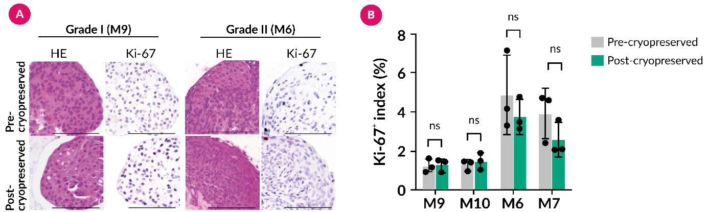
Figure 4. Post-thaw meningioma organoids retain histological features and proliferative activity. (A) Representative H&E sections show that the post-thaw organoids preserve a densely cellular, organized tissue architecture comparable with the pre-cryopreservation culture. Ki-67 staining identifies proliferating nuclei before freezing and in both post-thaw examples after one week of recovery. Scale bars: 100 μm. (B) The Ki-67-positive index measured across three fields shows no significant difference between conditions (mean ± SD; P = 0.543), indicating that proliferative capacity was maintained within the limits of the analysis. Image Credit: NIPPON Genetics EUROPE GmbH
For a three-dimensional model, these readouts determine whether the organoid is recovered as an organized, proliferative culture rather than simply as a collection of living cells.
iPSC-derived cardiomyocytes for electrophysiological risk stratification
In a study by Khudiakov et al., patient-specific hiPSC lines were differentiated into cardiomyocytes and subsequently cryopreserved in Bambanker™ between days 9 and 16 of differentiation.3 Following thawing, replating, and expansion, corrected field-potential duration was compared across wild-type, low-risk, and high-risk long-QT syndrome variants (Figure 5).
Repolarization values were longer in most high-risk variants than in wild-type or low-risk groups, despite distribution variations and overlap between variants. These data indicate that disease-relevant electrophysiological variations remained measurable following cryobanking and differentiation.
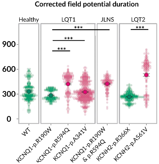
Figure 5. Recovered hiPSC-derived cardiomyocytes reproduce long-QT syndrome-associated electrical phenotypes. Corrected field-potential duration was measured by multielectrode array after cryobanked hiPSC lines were thawed, expanded, differentiated and replated as cardiomyocytes. Each distribution summarizes 157–547 measurements for a wild-type line or an LQTS variant. Green denotes wild-type or low-risk variants and pink denotes high-risk variants. Most high-risk variants show longer corrected field-potential durations than wild-type or low-risk groups, while the varying width and overlap of the distributions illustrate variant-specific heterogeneity. Kruskal–Wallis with Dunn’s test and Benjamini–Hochberg correction: *P ≤ 0.05, **P ≤ 0.01, ***P ≤ 0.001. The panel demonstrates preservation of disease-relevant electrophysiological readouts after the cryobanking workflow; it is not a fresh-versus-frozen comparison. Image Credit: NIPPON Genetics EUROPE GmbH
Cryobanked iPSCs for microglia-like cell models
In a study by Kalf et al., Bambanker™ was used to bank control and tuberous sclerosis complex (TSC) iPSC cultures prior to differentiation into induced microglia-like cells.4 The techniques report thawing the iPSCs in the presence of Y-27632 for 24 hours. The differentiated cells were subsequently assessed with microglial identity markers and functional assays that covered calcium signaling, mitochondrial respiration, inflammatory responses, and synaptosome uptake.
The resulting cells expressed microglial markers, including IBA1 and TMEM119 while retaining measurable disease biology. Compared with controls, TSC-derived microglia-like cells exhibited increased uptake of pHrodo-labelled human synaptosomes at both 1 and 24 hours, as well as alterations in calcium signaling and mitochondrial activity. These findings show that the cryobanked iPSC workflow progressed to lineage-specific differentiation and biologically informative functional modeling.
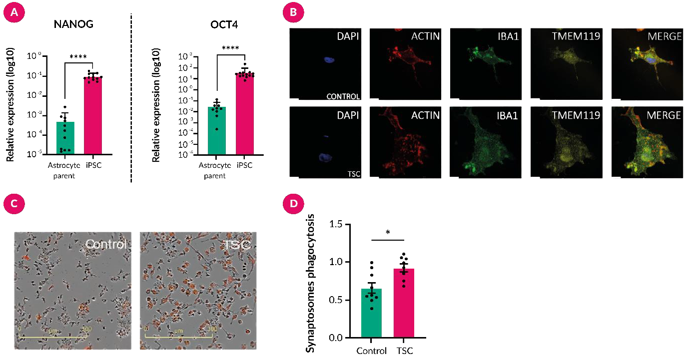
Figure 6. Bambanker-banked iPSCs support downstream microglial modeling. (A) NANOG/OCT4 expression after reprogramming. (B) ACTIN, IBA1 and TMEM119 staining in control and TSC microglia-like cells (DAPI; scale bar: 10 μm). (C,D) Representative pHrodo-synaptosome uptake and quantification after 24 h (scale bar: 300 μm; control, n = 4; TSC, n = 3; ≥ 2 experiments). Mean ± SEM; (A) ****P < 0.0001; (D) Mann–Whitney U test, *P < 0.05. Image Credit: NIPPON Genetics EUROPE GmbH
Defining recovery for a specific model
A robust post-thaw qualification plan begins with the downstream decision the model must support.
From there, a small set of staged measurements can be selected.
- Immediately after thawing. Viability and cell yield should be measured using a defined technique and time point. The thawing process, dilution stages, and use of recovery supplements should also be recorded.
- During early recovery. Attachment or aggregate reformation, morphology, outgrowth, and the time needed to resume growth should be evaluated. For three-dimensional models, size distribution and structural integrity should be included where appropriate.
- Following culture re-establishment. Model identity or lineage should be confirmed using suitable markers. Based on the system, this may include pluripotency, differentiation, cell-type composition, or histological organization.
- At the point of use. The functional readout relevant to the experiment should be executed, such as electrophysiology, secretion, contractility, barrier properties, uptake, response to a perturbation, or another confirmed endpoint.
Practical questions before freezing iPSC and 3D models
- Which material is being banked: undifferentiated iPSCs, differentiated cells, aggregates, organoids, or tissue fragments?
- Which recovery endpoint predicts whether the model is suitable for its intended experiment?
- When after thawing should each endpoint be assessed?
- Which fresh or established reference condition will be employed for comparison?
- Are aggregate size, cell density, freezing volume, cooling conditions, storage temperature, thaw rate, and recovery supplements controlled and documented?
References
- Hiramatsu, S., et al. (2022). Cryopreservation of induced pluripotent stem cell-derived dopaminergic neurospheres for clinical application. Journal of Parkinson’s disease, 12(3), pp.871–884. DOI:10.3233/JPD-212934. https://journals.sagepub.com/doi/10.3233/JPD-212934.
- Yamazaki, S., et al. (2021). Newly established patient-derived organoid model of intracranial meningioma. Neuro-oncology, 23(11), pp.1936–1948. DOI:10.1093/neuonc/noab155. https://academic.oup.com/neuro-oncology/article/23/11/1936/6313216?login=false.
- Khudiakov, A., et al. (2026). Machine learning-guided risk stratification for long QT syndrome genetic variants with hiPSC-derived cardiomyocytes. Cardiovascular Research, 122(10), pp.1374–1390. DOI:10.1093/cvr/cvag105. https://academic.oup.com/cardiovascres/article/122/10/1374/8678446.
- Kalf, R.S., et al. (2026). TSC-associated microglial hyperactivity: Enhanced calcium signaling, metabolism, and phagocytosis. Acta Neuropathologica, 151(1). DOI:10.1007/s00401-026-02986-8. https://link.springer.com/article/10.1007/s00401-026-02986-8.
About NIPPON Genetics EUROPE GmbH
Our contribution to your success – we are committed to ensuring that you can achieve your goals!
NIPPON Genetics EUROPE is a global biotech company founded in 2005 with its own research, development, and production based in Germany. By providing an extensive range of selected and groundbreaking products for the life science sector, we would like to be a reliable partner and contribute to achieving our customers' individual goals. The majority of our employees have a scientific background because we want to understand which problem solutions our customers care about.
Sponsored Content Policy: News-Medical.net publishes articles and related content that may be derived from sources where we have existing commercial relationships, provided such content adds value to the core editorial ethos of News-Medical.net, which is to educate and inform site visitors interested in medical research, science, medical devices, and treatments.