Whole-body low-dose computed tomography uses low doses of radiation to analyze the internal organs of an individual. This tool is useful in clinical medicine and research as it allows for the diagnosis and treatment of certain diseases.
Skip to

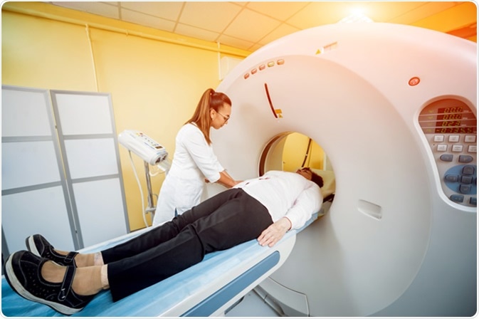
Doctor and patient in the room of computed tomography at hospital. Image Credit: Romaset / Shutterstock
What is computed tomography?
The most common computed tomography (CT) scanning method is X-ray CT. This works by taking many X-ray measurements from multiple angles and combining them to produce a cross-sectional image of a specific area within an object, which allows the inside of an object to be viewed. The most common use for CT scans is medical imaging, where cross-sectional images are used to diagnose diseases and help with the treatment planning.
CT scans are also extremely useful for research purposes, as it allows for whole organisms to be imaged, which can be particularly useful for developmental biology, where several images of the same organism can be taken over time. CT scans can be used to generate three-dimensional (3D) models of whole organisms and organs within larger organisms.
Other methods of CT scanning include magnetic resonance imaging (MRI), ultramicroscopy, and confocal laser scanning microscopy (CLSM). These methods also allow for high resolution imaging down to the sub-micrometer level. However, only X-ray CT and MRI can be used for larger tissues (> 500 μm), as they do not require complex sample preparation.
Low Dose CT Scans to Look for Lung Cancer
The effect of the radiation dose when using X-ray computed tomography
X-ray CT is considered a non-destructive imaging technique, as a sample or organism doesn’t need to be damaged in order to perform the imaging. An aspect of X-ray CT that isn't always initially considered is the effect that the X-ray dose could have on the subject. In this case, the dose is the amount of energy that is absorbed by an object after exposure to radiation, which is measured in gray (Gy).
This is especially true when performing X-ray CT scans in humans as short-term and long-term over-exposure to X-ray radiation can cause a multitude of problems. Exposure to approximately 2 Gy can cause mild symptoms, such as diarrhea, but immediate exposure to over 30 Gy can cause seizures, leukemia, or death.
What are some uses for whole-body low-dose computed tomography?
Whole-body low-dose computed tomography (WBLDCT) is an X-ray CT scanning method that uses a low dose of radiation to analyze the whole body of an individual. WBLDCT is a very versatile technique that can be used to diagnose and assess patients with illnesses such as cancer and bone fractures.
During one study, WBLDCT was compared with conventional radiography (CR) to assess which method had better diagnostic performance to detect bone lesions, skeletal fractures, vertebral compressions, and other extraskeletal findings. The radiation exposure for both methods was extremely small (approximately 2.5 mGy). The study discovered that WBLDCT was more accurate than CR as it detected more rib fractures, vertebral compressions, and other extraskeletal findings, and it had less incorrect estimations of the disease stage than CR.
The standard imaging tool for multiple myeloma (MM) is plain radiography, which delivers a high dose of radiation when used to scan the entire body. WBLDCT was tested to determine if it could effectively evaluate patients of MM. 41 patients with MM were analyzed with WBLDCT, which found that it had a higher sensitivity than plain radiography. The radiation dose during the WBLDCT analysis was approximately 7.4 mGy, which is significantly lower than the radiation dose of plain radiography. WBLDCT was much faster and more convenient for the patients than radiography.
Conclusion
Radiation dose is an important consideration when performing imaging for clinical diagnosis and medical research. WBLDCT may provide advantages over existing methods with respect to convenience and safety.
Further Reading
Last Updated: May 9, 2019