Introduction
How is flow cytometry used in anti-fungal research?
How is flow cytometry used to test atmospheric spores?
Flow cytometry and fungi in the food industry
References
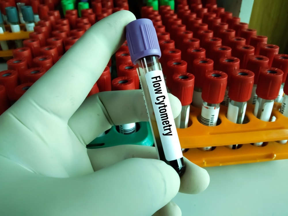
Flow cytometry is a powerful analytical tool used for high throughput particle or cell analysis, wherein the sample is passed in a single file through the incident light of a laser detector, allowing information about particle size and density to be inferred.
The technique is widely used in the pharmaceutical industry and in research, including the field of mycology, the study of fungi such as mushrooms and yeasts. Some of the key uses of flow cytometry in mycology will be discussed below.

Image Credit: Arif biswas/Shutterstock.com
How is flow cytometry used in anti-fungal research?
The influence of anti-fungal drugs and therapeutics against fungi and their spores is commonly assessed by flow cytometry in research, just in the same manner as eukaryotic cells or bacteria may be assessed in their sensitivity towards therapeutics or antibacterial drugs. Flow cytometry very frequently incorporates fluorescence excitation and detection apparatus that allows the quick identification of cellular features by the intensity of fluorescence once tagged. For example, a tag bound to an antibody specific to the cell mitochondria might be employed, allowing cells carrying more mitochondria to be distinguished.
Flow cytometry can be highly comparable to traditional viability testing when determining minimum inhibitory concentration, as demonstrated by Ramani et al. (2003) when comparing the efficacy of common anti-fungals against the three most common human pathogenic fungi: Aspergillus fumigatus, A. flavus, and A. niger. Viable cells were stained following incubation with different concentrations of the drugs and counted by flow cytometry; thus, the drug concentration required to inhibit cell growth determined by comparing cell counts.
A significant degree of additional quantitative information can be gained using flow cytometry by simultaneously staining fungal cells with a cocktail of fluorophores that indicate the relative proportions of each target, and the information relating to the mechanism of action of the therapeutic might be inferred. Further, flow cytometry is able to sort cells of interest for further examination, allowing the downstream effects of the anti-fungal to be investigated in detail.
How is flow cytometry used to test atmospheric spores?
Fungal spores make up most of the mass of biological aerosols in the atmosphere, and fungal contamination is a constant concern in sterile laboratory or industrial environments. In work and living environments fungal spores may be detrimental to human health if excessively inhaled, and they also play a role in the environment and cloud formation by acting as nuclei for the generation of ice crystals.
The detection and quantification of fungal spores in the atmosphere is therefore useful to multiple industrial and research applications, though unfortunately the vast majority of airborne spores cannot easily be cultured, particularly when mixed in with much more abundant or quickly growing fungi. Of those that can be cultured they must then be assessed by light microscopy, which is time consuming and potentially subjective.
Flow cytometry is therefore extremely useful in the high throughput analysis of fungal spores, where the various biological microorganisms can be stained with a number of fluorophores for quick identification. Where fluorescent staining of the dozens of varieties of fungal spores and other biological entities in an atmospheric sample may be impractical flow cytometry can also be tandem coupled with analytical methods such as gas chromatography-mass spectrometry to allow quantification of the constituents.
Gas chromatography effectively separates the constituent molecular components of a sample for improved resolution, and mass spectrometry quantitatively indicates the molecular content of the sample. Particular molecules may be abundant to certain microorganisms, and thus their molecular components can be used to quantify their presence. For example, mannitol and arabitol sugars are particularly common in fungal spores, and so quantifying their presence can allow the number of fungal spores to be determined. Ambient fungal spores can be collected for flow cytometry by pump vacuum and then exchanged into a liquid medium, whereupon they can be stained prior to analysis.

Image Credit: Pixel-Shot/Shutterstock.com
Flow cytometry and fungi in the food industry
Flow cytometry is used in the food industry to regulate and characterize yeast where it is employed, for example when brewing beer or during other fermentation processes. Ensuring that the correct number of yeast cells are in culture and that they are growing at the correct rate is vital to successful fermentation, otherwise generating non-optimal growth conditions, affecting taste and causing issues with filtration.
Culture based methods are too slow to be useful during industrial processes, and so if flow cytometry is not available yeast cells are dyed with methylene blue to distinguish living from dead cells and assessed by light microscopy. Only a comparatively low number of cells can be analyzed in this way, and results may be subjective to the observer, and thus flow cytometry is extremely useful in the high throughput assessment of culture viability. Flow cytometry cannot easily detect cells colored with methylene blue, and thus dead cells are instead stained with fluorescent oxonol dye.
A culture sample can be stained with multiple fluorescent dyes to allow simultaneous detection of bacterial contamination and analysis of other parameters, including the stage of the cell cycle the yeast is currently in.
Stage cycle is indicated by the quantity and spread of DNA within the cell, and thus fluorescent tags to yeast DNA are employed, where elongation indicates that the cell is entering the M phase and separation of DNA indicates the S phase. It is useful to understand the cell cycle state and rate of progression in yeast cells during fermentation as it is in the G1 phase that most RNA and DNA synthesis takes place and cell budding occurs, producing more bitterness and fermentation activity.
References:
- Boyd, A. R., Gunasekera, T. S., Attfield, P. V., Simic, K., Vincent, S. F., & Veal, D. A.. (2003). A flow-cytometric method for determination of yeast viability and cell number in a brewery. FEMS Yeast Research, 3(1), 11–16. https://doi.org/10.1111/j.1567-1364.2003.tb00133.x https://www.sciencedirect.com/science/article/pii/S0021850213001845
- Liang, L. et al. (2013) Rapid detection and quantification of fungal spores in the urban atmosphere by flow cytometry. Journal of Aerosol Science, 66 https://www.ncbi.nlm.nih.gov/pmc/articles/PMC368332/
- Prigione, V., Lingua, G., & Marchisio, V. F.. (2004). Development and Use of Flow Cytometry for Detection of Airborne Fungi. Applied and Environmental Microbiology, 70(3), 1360–1365. https://doi.org/10.1128/aem.70.3.1360-1365.2004 https://www.ncbi.nlm.nih.gov/pmc/articles/PMC253779/
- Ramani, R., Gangwar, M., & Chaturvedi, V.. (2003). Flow Cytometry Antifungal Susceptibility Testing of Aspergillus fumigatus and Comparison of Mode of Action of Voriconazole vis-à-vis Amphotericin B and Itraconazole. Antimicrobial Agents and Chemotherapy, 47(11), 3627–3629. https://doi.org/10.1128/aac.47.11.3627-3629.2003 https://journals.plos.org/plosone/article?id=10.1371/journal.pone.0017175
- Knutsen, J. H. J., Rein, I. D., Rothe, C., Stokke, T., Grallert, B., & Boye, E.. (2011). Cell-Cycle Analysis of Fission Yeast Cells by Flow Cytometry. PLOS ONE, 6(2), e17175. https://doi.org/10.1371/journal.pone.0017175
Further Reading
Last Updated: Feb 3, 2023