In this interview, NewsMedical speaks with Arvonn Tully and Esther Kieserman from Yokogawa Life Science about how confocal-based high-content imaging is advancing core facility research and improving data reliability.
Could you introduce yourselves and share your background, particularly your work with confocal-based high-content imaging and analysis?
Esther Kieserman (EK): I have worked in the microscopy industry all throughout my career, starting back in my sophomore year of college. I earned a Ph.D. in molecular biology focusing on confocal-based imaging, and my postdoctoral work primarily involved widefield microscopy.
I then spent approximately three years working in a microscopy core facility, during which time I transitioned from being a microscope user to a microscopist. Before joining Yokogawa Life Science's team in February 2024, I would say that I had limited direct experience with high-content imaging.
I spent about five years working with traditional microscopes, followed by four years focused on training, application support, and technical assistance. Microscopy has always been a constant in my career, and high-content imaging represents a unique, specialized branch of this field. My background in traditional microscopy gives me a fresh and well-rounded perspective when approaching confocal-based high-content imaging and analysis.
Arvonn Tully (AT): I have been part of Yokogawa’s High Content Analysis team since May 2020. Before that, I spent 14 years working with 3D image analysis software in the research microscopy field, collaborating closely with the Imaris and Arivis teams across North America. During this time, I supported labs, core facilities, biotech, and pharmaceutical companies, both before and after sales, helping them get the most out of their imaging tools.
What I enjoy most is helping researchers turn their ideas into meaningful 3D measurements. My background in scientific research has been invaluable as I have worked to bring high-content analysis into 3D environments, particularly in drug discovery.
I am especially passionate about using models like spheroids, organoids, and patient-derived tissues to create more realistic and physiologically relevant studies. Confocal microscopy, with its ability to optically section through 3D cultures, plays a key role in exploring the behavior of cells within these complex systems, and I am thrilled to be part of such innovative work.

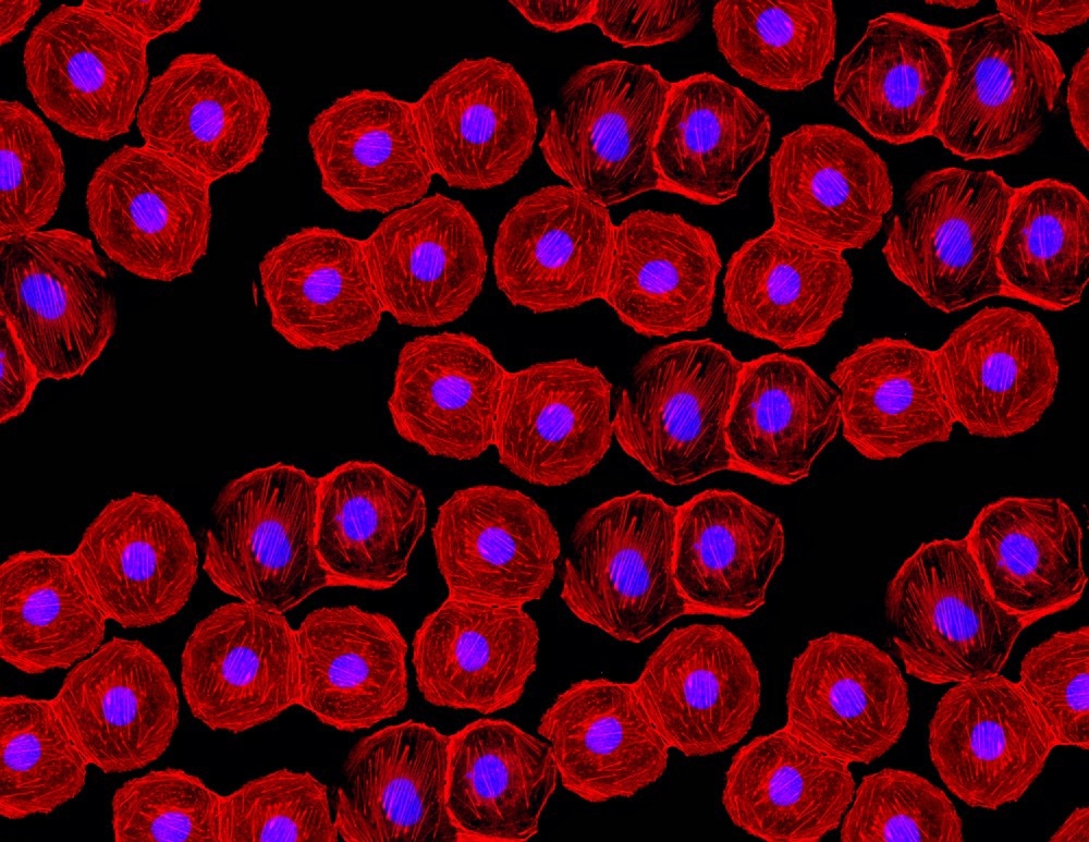
Image Credit: Ellen Curtis/Shutterstock.com
Esther, from your experience in managing core facilities, what are the primary challenges these facilities face regarding resource allocation, data handling, and throughput?
EK: A primary challenge for core facilities is acquiring sufficient resources to invest in new technology or having the foresight to keep pace with technological advancements.
Core directors must invest in "future-proofed" equipment to ensure their facilities remain attractive to both the institution’s researchers and the new researchers they are trying to recruit.
The funding cycle, at least in the United States, is long. Core facility managers must make decisions months, if not years, in advance of placing a piece of equipment in their facility. This can significantly strain the core and impact its ability to secure further funding if the equipment choice is incorrect and remains unused.
Data management has consistently posed issues for core facilities. Ten years ago, we used external servers with time limitations on local data storage. This challenge has since intensified as data production speeds have increased and researchers generate more data per experiment.
Compounding this issue is the growing size of data files, which demand more computer storage. We frequently encounter situations where a single overnight experiment can occupy an entire multi-terabyte hard drive.
Throughput is also a major concern. Researchers in core facilities typically pay hourly to use equipment, whether it be a flow cytometer, a confocal microscope, or even a standard compound microscope. Costs rise if specialized assistance is required.
The price also increases if a researcher needs a core manager to conduct a flow cytometry run, image brain slices on the confocal, or analyze a sample through mass spectrometry. Researchers and industry experts aim to maximize the data they obtain from their investments; thus, any equipment downtime can be detrimental to project progress.
How do you see confocal-based high-content imaging addressing some of these challenges, particularly in streamlining workflows and enhancing data reliability?
EK: Confocal-based high-content imaging can address several core facility challenges, especially in terms of throughput. High-content imaging and analysis alleviate strain on more specialized equipment.
While not a direct replacement for flow cytometry, a high-content system can serve as a quick alternative for a panel of up to four colors, offering a convenient way to count cell populations without needing to spend time dissociating cells into suspension.
Instead of utilizing an expensive point-scanning confocal microscope, researchers can achieve a similar quantity and quality of images using a spinning disk-based high-content system within the same timeframe.
In many cases, high-content analysis systems are a direct replacement or even an improvement over traditional methods.
Once the parameters are defined, high-content imagers consistently execute experiments with precision, ensuring reliability and reducing the influence of biased analysis routines. This consistency not only enhances the accuracy of results but also eases the mental burden on researchers. With less time spent managing repetitive tasks, researchers can focus on asking innovative questions, exploring creative ideas, and advancing their projects.
The reliability of data is a major advantage, allowing researchers to concentrate on experimental design without the added concern of whether the experiment will run smoothly. This frees up their time and energy to focus on other important tasks within the lab or facility.
Arvonn, given your expertise in software analysis, can you elaborate on the role of software in making confocal-based high-content imaging a valuable tool for core facilities?
AT: Automation advancements are a game-changer for core facilities, making it easier to get researchers up to speed and significantly boosting the reliability of research data. High-content imagers, for instance, allow scientists to gather meaningful results much faster than traditional microscopes ever could.
These microscopes are fully automated and user-friendly, operating more like plug-and-play devices. Their defined workflows not only simplify tasks but also minimize training time and reduce the likelihood of user errors.
In today's research landscape, automation is essential for scaling experiments. The bar for publishing papers has risen—experiments now require much larger sample sizes to confirm the validity of results, with ten samples no longer cutting it. High-content confocal microscopes rise to this challenge with advanced spinning disk technology.
Unlike traditional laser point-scanning confocals, they use 1,000 light beams simultaneously to capture images more quickly and with greater sensitivity. This speed and precision are vital for producing high-quality data.
How does implementing confocal-based high-content imaging differ between industrial and academic core facilities? Have you observed unique challenges or advantages?
EK: Industrial core facilities are often among the first to adopt high-content imaging systems, typically investing in equipment tailored to specific projects and operating it continuously for those purposes. For industrial users, repeatability and minimal downtime are crucial, as experiments often run 24/7. In these settings, ease of use becomes a major advantage, especially since troubleshooting during off-hours can be challenging. These systems need to perform reliably every time they are used.
In contrast, academic core facilities tend to prioritize versatility, seeking equipment that is both user-friendly and adaptable to a variety of applications. For instance, a confocal high-content system can easily be adjusted to handle tasks as varied as scanning 3D live cells and imaging brain tissue slices.
Do you find that data analysis requirements differ between industrial and academic settings?
AT: Analysis in industrial settings generally follows one of two approaches: targeted or comprehensive.
Targeted groups use well-characterized assays that address specific questions about cell populations. The other approach involves measuring everything and using deep learning tools to identify distinct phenotypes and effects in secondary data analysis.
As core facilities evolve, how does high-content imaging integrate with other core technologies like flow cytometry or genomics? Do you foresee a trend toward a multimodal approach?
EK: High-content imaging is increasingly being adopted in non-traditional imaging cores, especially flow cytometry.
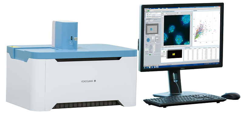
Systems like the CQ1, which can save data in FCS format, offer researchers the flexibility to work with various dyes and collect substantial data from images. While it does not fully replace an 18- or 20-dye flow cytometer panel, it provides a valuable starting point for comprehensive flow cytometry experiments.

Image Credit: Yokogawa
One major advantage of high-content imaging is its ability to streamline simpler flow experiments. Researchers can skip steps like removing cells from plates or transferring them into flow solutions or machinery. Instead, they can go straight from incubation to data collection, preserving time-based data that is often lost in traditional flow cytometry processes.
Confocal high-content imaging and flow cytometry are on track to become industry-standard multimodal technologies. There is also increasing interest in combining confocal imaging with mass spectrometry cores. This integration offers a unique ability to merge spatial and temporal data from imaging with mass spectrometry analysis, delivering deeper insights into cell-to-cell heterogeneity—critical for advancing disease research and treatment strategies.
Could you share some insights on managing high-volume image data? What practices do you recommend for core facilities to streamline data storage, analysis, and accessibility?
AT: Effective data storage policies are critical for core facilities, ensuring clear guidelines on retention periods and backup options to address user errors. While storage needs vary, many publications require raw data to be stored for up to seven years. Collaborating with users to develop storage and sharing solutions is key, with tools like Omero often used to manage metadata and analysis results.
Cloud storage is another option, though it can be prohibitively expensive for individual labs. Some forward-thinking labs are adopting multi-petabyte storage solutions within a single on-site rack. This setup supports internal access while aligning with FAIR standards, providing a path for broader data sharing with the research community.
Data analysis tools also involve trade-offs. Open-source options are cost-effective and customizable but may require additional training and support, making their overall cost comparable to commercial software, which provides more comprehensive support. Facilities must weigh these factors to select the best tools for their needs.
How can confocal-based high-content imaging and advanced analytics improve the overall experience for researchers and technicians within core facilities?
AT: High-content imaging systems streamline workflows, optimizing them to address specific assay questions at scale. Features like automatic batch processing and integration with scheduling software enable researchers to run large-scale experiments with minimal time spent at the microscope. Automated data transfer and compatibility with third-party analysis tools further boost efficiency, allowing researchers to focus on insights rather than logistics.
Looking forward, what improvements would you like to see in high-content imaging technology and analysis software to enhance the capabilities of core facilities?
EK: From my experience in microscopy, flexibility, upgradability, and futureproofing are crucial for core facilities. Managers want assurance that their investment will remain functional and attractive for at least five years.
High-content systems should become more upgradeable without sacrificing their ease of use. Innovations like these will help core facilities offer better services, improve data quality, and support cutting-edge research across various disciplines.
AT: The next major step is an increased focus on 3D spatial biology. Research has shown that 3D cellular biology differs significantly from 2D monolayers. The research community has likely reached the limit of 2D studies, and it is time to explore 3D interactions of cells in physiologically relevant environments.
Some questions can only be addressed through 3D live cell models and fixed tissue, which could revolutionize pharmacology and personalized cell therapies. It really is an exciting time for the field.
About Arvonn Tully
Arvonn Tully is a 3D image analysis expert with over 15 years of experience developing innovative solutions for complex biological challenges. In particular he is interested in sub cellular tracking, neuronal reconstruction, and finding solutions for complex analysis problems. Prior to Yokogawa, Arvonn worked at other image analysis companies specializing in 3d visualization and analysis of large multi-dimensional images. He trained thousands of users in 3d Image analysis and developed numerous novel 3d analysis solutions. In particular he developed a bouton finder, and a novel solution to identifying and tracking the contact area in 3d between touching endosomes. He started his career in biological research at Dr. Levitan's Lab in University of Pittsburgh Medical College. There he used several imaging techniques to study the biophysics of large dense core vesicle mobility using D. melanogaster neuromuscular junctions. Originally from Virginia, he grew up in the mountains and playing in the halls of Virginia Tech.
About Esther Kieserman
Dr. Esther Kieserman is a skilled microscopist with over 20 years of experience specializing in confocal-based imaging and development of innovative microscopy techniques. She earned her undergraduate degree at Carnegie Mellon University and went on to earn a Ph.D. in molecular biology in the lab of Dr. John Wallingford at UT Austin. Esther completed her postdoctoral research in the lab of Dr. Rebecca Heald at UC Berkeley. Before joining Yokogawa Life Science in February 2024 as Head of 3rd Party Collaborations and Marketing Manager, Esther spent several years at Johns Hopkins University’s microscopy core facility and nine years at Nikon Instruments, where she worked on sales, training, application support, and technical assistance. Originally from upstate New York, Esther is an avid animal lover and enjoys caring for her rabbits. With a rich background spanning research, technical support, and commercial applications, she brings a well-rounded and comprehensive perspective to the field of high-content microscopy.
About Yokogawa Life Science

We have 30 years of experience in this life science field and will respond to customer's problem solving with cutting-edge solutions.
Our confocal scanner unit CSU series enables 3D observation of the cells in detail and dynamics of organelles inside cells. Since the CSU series is capable of high-speed shooting, it is also suitable for observing high-speed life phenomena. In addition, the CSU series is a multi-point confocal method which is extremely gentle to cells, best suitable for long-term live cell observation.