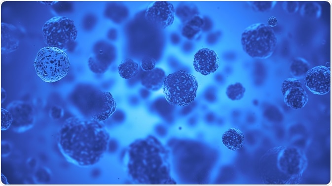
DAPI, or 4,6-diamidino2-phenylindole, is a fluorescent nucleic acid dye used to preferentially stain double-stranded DNA.
There is some evidence that it preferentially binds AT clusters present in the minor groove of the DNA.
 Image Credit: Alexey Godzenko / Shutterstock
Why use DAPI?
Other nuclear stains used to visualize DNA during interphase and mitosis include Hoechst stain, ethidium bromide, propidium iodide and acridine orange.
However, DAPI is preferable because it is more photostable than the other dyes. DAPI specifically stains double stranded DNA with no non-specific labeling in the cytoplasm.
For DAPI to enter cells and bind to DNA, the cells need to be permeabilized. After binding to DNA, the fluorescence of DAPI increases 20 fold.
Preparing the DAPI stock solution
To the start the staining process, first a stock solution of DAPI needs to be prepared. For this, the contents of a vial of DAPI (10mg) are dissolved in 2mL of dimethylformamide (DMF) or deoinised water.
As DAPI is not very water-soluble, this process may take some time and may require additional methods, such as sonication.
DAPI is also not very soluble in phosphate buffer saline (PBS).
For storage, DAPI solution can be kept at 2−6°C for a short time, but to store the solution for longer durations it should be kept at ≤-20°C.
The entire process should be performed using gloves and should be performed carefully as DAPI is a known mutagen.
Fluorescence characteristics of DAPI
The excitation wavelength for DAPI is 358 nm, while the emission wavelength is 461 nm. Xenon, mercury-arc lamp, or UV light can be used to excite DAPI during experiments.
DAPI can be used to stain nuclei using fluorescent microscopy, flow cytometry, or to stain chromosomes in fluorescence in situ hybridization (FISH).
DAPI staining for fluorescence microscopy
Microscopy is a method for visualizing small things, such as cells, and fluorescence can be added to microscopy to see certain features of cells, like the nucleus using DAPI.
First the sample is fixed using an appropriate fixation protocol. After fixing, the sample is equilibrated with PBS. The DAPI is then diluted and added to the sample so that the sample is completely covered.
This is then incubated for around 5 minutes, and then rinsed in PBS few times to remove excess stain and buffer from the sample. Reducing the number of washes can lead to non-specific stain.
In the end, a mounting medium with an antifade reagent can be used to mount the sample onto the coverslip.
Antifade reagents reduce photobleaching and thus, help in preservation of the fluorescent signals for a long time. The sample can then be visualized using a fluorescent microscope.
DAPI staining for flow cytometry
Flow cytometry is a technique which is used to identify different cells within a mixed population of cells. The methodology for DAPI in flow cytometry is given below:
- First, the sample can be fixed using the appropriate fixation protocol specific to the sample.
- A suspension of f 2 × 10⁵ to 1 × 10⁶ cells should be taken and these cells are pelleted using centrifugation.
- The pelleted cells are resuspended at room temperature in PBS.
- The resuspended cells are then transferred to absolute ethanol at -20°C for 15 minutes.
- After this step, the cells are pelted again using centrifuge and resuspended in PBS.
- For the staining, add the diluted DAPI stock solution to pelleted cells.
- After incubation for 15 minutes, flow cytometry can be performed in the presence of the dye.
DAPI staining for FISH (Fluorescence In Situ Hybridisation)
FISH is a method used to visualize chromosomes using fluorescence microscopy. The methodology used in FISH is given below:
- The sample should be fixed appropriately and rinsed in deionized water to remove residual buffer and non-specific staining.
- Add the diluted DAPI stock solution on to the sample.
- After incubating in DAPI for 30 minutes, the coverslip can be removed and washed with PBS or deoinised water to remove excess DAPI.
- Excess liquid can be further removed using blotting paper.
- The coverslip should placed on the slide and edges sealed using wax or polish.
- Finally, the sample can be visualised using a fluorescence microscope.
Reducing autoflourescence
Some cellular molecules, such as nicotinamide adenine dinucleotide or flavin adenine dinucleotide can auto-fluoresce, or exhibit fluorescence naturally.
This can reduce the signal to noise ratio when using fluorescent stains such as DAPI, which can make clear visualization and interpretation of images difficult.
This phenomenon is seen more in instances where the excitation wavelength of auto-fluorescing molecules (such as nicotinamide adenine dinucleotide) and target molecules (DNA) are similar.
This can be overcome by the addition of reducing agents or imaging unlabeled control slides, which would show auto-fluorescence but not the specific fluorescence of target molecules.
Further Reading
Last Updated: Oct 1, 2018