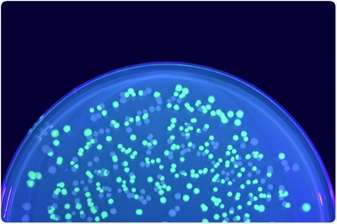
Single-particle tracking (SPT) is a rapidly developing technique in cellular biology. This technique has been used to define the diffusion dynamics of nanoparticles and biomolecules, through the use of fluorescent probes and fluorescent microscopy.
 Image Credit: KPWangkanont / Shutterstock
Principle
In SPT, the probe is attached to the molecule of interest, such as an organelle, virus, or a receptor to view the individual characteristics of the molecule.
Molecules can interact stochastically with other molecules or cellular structures, which helps in distinguishing the various stochastic processes of a molecule.
SPT tracking methods
Particle detection can be carried out by the manual and automatic tracking method. The manual method is labor intensive and is sometimes difficult where large datasets are required.
In this situation, SPT software programs are used to generate automated algorithms for detecting and linking a particle. The different fluorescent probes that are used in SPT are described below.
Dual color single particle tracking via nanobodies
Quantum dots
Quantum dots are considered efficient fluorescent probes as they are more photostable than other organic dyes and probes. This makes them an ideal choice for long-term SPT experiments and live cell applications.
Fluorescent proteins
Though fluorescent proteins are made highly specific by genetic engineering, they are bigger and less photostable compared to organic small-molecule fluorophores.
Organic small-molecule fluorophores
Organic small-molecule fluorophores are prepared by chemical labeling of proteins or by physically incorporating them into the target molecule.
Gold nanoparticles
The gold nanoparticles have a small particle size ranging from two to 100 nm and show no cytotoxicity. They display strong light scattering properties at the plasmon resonance wavelength.
Other fluorescent probes
Apart from these, many fluorescent probes have been developed using nanoparticles. An example of such a probe is single-walled carbon nanotubes (SWNT).
SWNTs are more stable (chemically and photophysically) than organic fluorophores. Another probe is the fluorescent nanodiamond (FND) that is extremely usefully for in vivo imaging.
The advantage of FND is that they are nontoxic and photostable and their surfaces can be modified to add different functional groups to expand their application over a wide spectral region.
Tracking procedure in SPT
The main advantage of quantum dots, gold nanoparticles, and FNDs are that they are resistant to photobleaching.
The tracking procedure involves the detection of single particles and reconstruction of the particle trajectory at each frame for quantification of the molecular movement. The important parameters are the diffusion coefficient time, the confinement time, and dwelling time.
Limitations of SPT
Some of the issues seen during the reconstruction of the particle trajectory are the appearance of a single trajectory as fragmented trajectories and the detection of other probes on non-transfected cells.
However, these problems can be limited by using probes of low particle density and partial manual algorithms for reconnection.
Applications of SPT
Analyzing cellular activity
SPT has been used to understand various activities in the living cells such as the therapeutic action of anti-cancer drugs, drug delivery, single-virus tracking, the study of mRNA, the study of endocytosis and exocytosis, and the study of membrane receptor transport.
For example, SPT has been used successfully in understanding the pathway and mechanism of transport of adrenergic receptors in real time in single living cells.
Virology
They have also been used successfully to study various viruses such as HIV and Dengue. Using real-time fluorescent microscopy and lipophilic fluorescent probes, it was demonstrated that the Dengue virus entered the cells through clathrin-mediated endocytic trafficking.
Further Reading
Last Updated: Dec 22, 2022