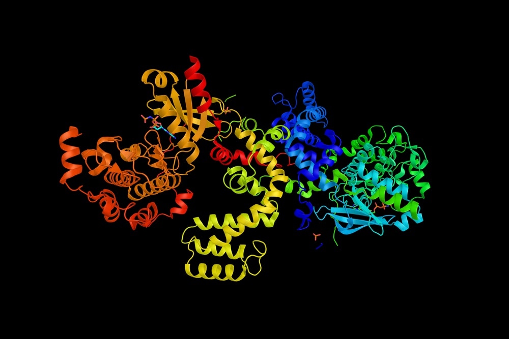
A review, by Professors Quast and Margeat from the French National Centre for Scientific Research exploring, has addressed G protein-coupled receptor (GPCR) structural dynamics and the role of single-molecule fluorescence SMF methods in their interrogation.
 Image Credit: ibreakstock / Shutterstock.com
The team focuses on single-molecule Förster resonance energy transfer (smFRET) in particular, which enables distances and, therefore, conformational changes to be measured across the timescales over which protein conformational movements occur. Most notably, biological insights into the activation and signaling for β2 adrenergic and the metabotropic glutamate receptors are unearthed.
G protein-coupled receptors (GPCRs) are highly physiologically relevant, and as such, are targets of pharmacological manipulation. Insights into their structural flexibility reveal the role of ligand-based activation.
Previous implications of this structural flexibility in physiology and pharmacology remain unexplored; this is particularly important, considering its potential in receptor fine-tuning and signal transduction specificity. Single-molecule fluorescence (SMF) offers a real-time, minimally structural invasive means of monitoring structural dynamics in the context of biological environments.
The timescales of protein fluctuations are capable of being measured at the single-molecule level. Fluorescent probes are excellent reporters of these conformational changes and can be used in living cells.
Using an ideal model, two states, separated by an energy barrier, are differentiated from one another either by plotting fluctuations (movements) in the fluorescently tagged protein area as a function of time or the plotting the distribution of the states. Through analyzing the dwell times (static periods, over which there are no movements), the tagged area spends in each of the states enables the rate of transition between each state to be determined.
However, molecular motions in proteins often occur faster than the time resolution of a single molecule fluorescence experiment, resulting in averaged conformations that prevent the dissolution of defined states.
Single molecules are observed by total internal reflection (TIRF), where changes in fluorescence properties per molecule as a function of time can be seen. Alternatively, diffusion-based measurements are used for fast protein dynamics as it offers excellent time resolution
Quast & Margeat, in their review, report the findings of the intracellular domain motions observed in class A GPCRs, and opening and closing and reorientation movements associated with the activation of the class C GPCR extracellular domain. These are all based on single-molecule fluorescence (SMF).
Several studies deducing the complex conformational landscape of the β2AR at the single-molecule level have been determined. They have also determined the effect of agonist binding on the dwell times of conformational states over milliseconds to seconds. The stabilization of agonist binding has pointed at the instability of the fully activated conformational state, which has been verified by other structural means.
Other receptor conformations have been subsequently determined. In particular, TMs 4 and 6 are known to undergo dynamic changes in motion upon ligand binding in a manner that depends on the ligand identity; they drive the efficacy of shifts in the rate at which the ligand-bound G protein-receptor complex is formed and also the efficiency of nucleotide exchange.
This latter finding represented a benchmark in the understanding of GPCR conformational dynamics; however, the authors note that complementary studies at higher time resolution are required for rates of interconversion between conformational states to be determined.
The authors also note research which labels GPCRs at alternative positions to determine concerted structural changes between the receptor and transducer. Namely, selective modification of amino acids: platelet-activating factor receptors (PAFRs), labeled on specifically incorporated azido-lyzines (AzK).
Single-molecule studies have also been used in the study of the extracellular domain (ECD) of class C GPCRs. Crystal structures of isolated ECDs from the mGluR2 using robust and highly specific labeling strategies (SNAP, HALO, and CLIP tags N-terminally fused to the receptors) have enabled elucidation of agonist-induced ECD dimer reorientation.
A pioneering study by Olofsson et al. revealed submillisecond structural dynamics of isolated mGluR2 extracellular domain dimers observed in freely diffusing molecules, showing the presence of two rapidly interchanging states. The same holds for members of all mGluR subgroups, which evidenced a general activation mechanism conserved across all mGluR subtypes.
A final study the authors note is by Vafabakhsh et al., who determined the conformational dynamics on surface-immobilized full-length mGluR2 using smFRET. These experiments were performed at a much slower time resolution and revealed a novel intermediate FRET population which led the researchers to conclude that the resting and active ligand-binding domain (LBD) may be stabilized by the TMD of the full-length receptors.
The β2AR and the mGluR receptors have been studied in-depth by single-molecule fluorescence studies, and as such, represent prototypical models for the class-A and class-C GPCRs. Altogether, the SMF observations on these receptors demonstrate the high degree of conformational flexibility within the receptor.
Most interestingly, partial agonists have been shown to work as poorly efficient modifiers of the conformational ensemble as opposed to stabilizers of distinct, partially active states. Quast and Margeat suggest that the future direction of SMF studies should be extended to verify if these same observations hold for other GPCRs.
Following this, SMF is hoped to be used to determine interactions with other partners. This will be useful in understanding ‘biased agonism’ – whereby pharmacological effects of ligands are determined by their ability to favor certain conformational states, leading to differential activity with their partners.
As technology develops, the ability to use non-invasively labels at potentially any desired position within the receptors and its ligands will enable experimental fine-tuning for further probing of conformational change with accuracy.
Finally, the authors predict smFRET experimentation in liposomes, nanodisks, and eventually, live cells will be common, owing to the effect that the membrane environment has on the conformational transitions.
Acknowledgments
The research is supported by a grant from the Agence Nationale pour la Recherche (ANR-12-BSV2-0015). The laboratory belongs to the France-BioImaging national infrastructure supported by the Agence Nationale pour la Recherche (ANR-10-INBS-04, “Investments for the future”
References
Olofsson, L. et al. Fine tuning of sub-millisecond conformational dynamics controls metabotropic glutamate receptors agonist efficacy. Nat. Commun. doi: https://doi.org/10.1038/ncomms6206 (2014).
Vafabakhsh, R. et al. Conformational dynamics of a class C G-protein-coupled receptor. Nature. doi:
https://doi.org/10.1038/nature14679 (2015)
Gregorio, G. et al. Single-molecule analysis of ligand efficacy in β2AR–G-protein activation. Nature. doi: https://doi.org/10.1038/nature22354 (2017)
Quast, R. B. & Margeat, E. Studying GPCR conformational dynamics by single molecule fluorescence. Molecular and Cellular Endocrinology. doi: https://doi.org/10.1016/j.mce.2019.110469 (2019)
Further Reading
Last Updated: Dec 2, 2019