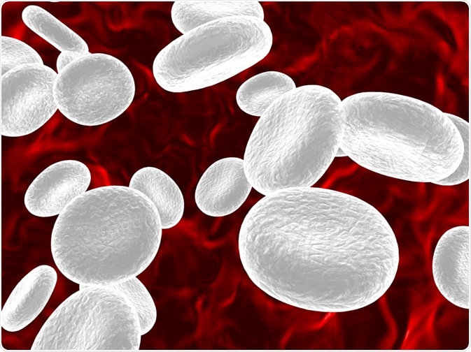
The use of flow cytometry in hematological research has grown immensely over the past several years. This analytical technique is capable of providing both qualitative and quantitative information about the surface proteins and glycoproteins present on erythrocytes, leukocytes and platelets.

Sebastian Tomus | Shutterstock
Flow cytometry and erythrocyte analysis
Rhesus Disease
One of the most widely used applications of flow cytometry is the analysis of fetal red cells present within maternal blood. In particular, flow cytometry is often used to diagnose Rhesus (Rh) disease in pregnant women, which can be detected as a Rhesis (Rh) antigen.
Rh antigens are expressed as a part of a protein complex on the membrane of red blood cells (RBCs). They are believed to play a role in maintaining the integrity of the RBC membrane.
To detect Rhesus disease, fluorescently labeled antibodies to the Rh antigen, as well as those that are directed against hemoglobin F, can be combined with flow cytometry. This method is technically superior to the traditional Kleihauer-Betke test used for Rhesus disease diagnosis, as it is more precise and sensitive in its ability to distinguish fetal cells from adult red cells that display small amounts of hemoglobin F.
Evaluating RBC Shape
The red blood cell membrane plays a crucial role in cellular deformation, a process that is essential for the exchange of oxygen within mature RBCs.
This processis influenced by a variety of cellular characteristics, including cellular geometry and cytoplasmic viscosity. Diseases such as diabetes, terminal renal failure, sepsis and acute inflammatory states cause RBCs to exhibit a more spherical shape as compared to normal disc-shaped RBCs.
Flow cytometry is useful here, as it can detect any shape alterations in RBCs to identify disease mechanisms and the likelihood of a patient developing a particular condition.
For example, flow cytometry used to categorize spherical RBCs can determine whether diabetes patients are more susceptible to diabetic microvascular conditions, such as diabetic nephropathy. For RBC shape analysis, flow cytometry is a reliable, rapid and highly reproducible technique.
CytoDiff and WBC Analysis
CytoDiff is a novel flow cytometric differential counting method that utilizes a 5-color/6-antibody reagent cocktail and an auto-gating algorithm to differentiate 18 white blood cell (WBC) populations, of which can include blasts, immunoglobulins (IGs) and lymphocyte subsets.
With the ability to count approximately 20,000 WBCs, CytoDiff provides significantly more precise cell counting abilities compared to manual cell counting processes are used. It is a highly sensitive, specific and rapid technique that has been clinically applied for the diagnosis and monitoring of leukopenia, sepsis or metastatic cancer.
Platelet analysis
Platelet analysis by flow cytometry can be achieved through both direct or indirect platelet-associated immunoglobulin assays. Flow cytometry plays a crucial role in the diagnosis of immune thrombocytopenic purpura (ITP), which is a genetic disorder that causes platelet consumption to be accelerated.
Unfortunately, since platelets are relatively small, uncolored and low in quantity compared to other blood cell populations, they are often difficult to count and characterize, especially in ITP patients that already exhibit lower platelet counts.
For this reason, flow cytometry has been found to be a sensitive method for platelet counting, as well as for the detection of platelet-associated immunoglobulin G (PAIgm), which is a marker for children with ITP.
Conclusion
Regardless of the sample type, flow cytometry provides a useful way for clinicians to rapidly measure intracellular characteristics for the diagnosis of various disease states, as well as monitoring the efficacy of treatments for these conditions.
Sources
- Brown, M., & Wittwer, C. (2000). Flow Cytometry: Principles and Clinical Applications in Hematology. Clinical Chemistry 46(8); 1221-1229.
- Dean L. Blood Groups and Red Cell Antigens. National Center for Biotechnology Information (US); 2005. Chapter 7, The Rh blood group.
- Piagnerelli, M., Boudjeltia, K. Z., Brohee, D., Vereerstraetten, A., Piro, P., et al. (2007). Assessment of erythrocyte shape by flow cytometry techniques. Journal of Clinical Pathology 60(5); 549-554.
- Kahng, J., Kim, Y., Kim, M., Oh, E., Park, Y., & Han, K. (2015). Flow Cytometric White Blood Cell Differential Using CytoDiff is Excellent for Counting Blasts. Annals of Laboratory Medicine 35(1); 28-34.
- Aref, S., Selim, T., Ibrahim, L., Abd-Elghaffar, H., & El Ashery, R. (2009). Flow cytometry detection of platelets autoantibodies in children with idiopathic thrombocytopenic purpura. Indian Journal of Hematology and Blood Transfusion 25(3); 96-103.
Further Reading
Last Updated: Jun 28, 2019