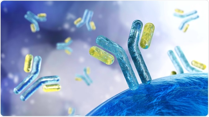
Antibodies that specifically react with self-antigens are called autoantibodies. These antibodies are generated as a result of the loss of tolerance response against self-antigens and can be pathogenic.
 Image Credit: ustas7777777/Shutterstock.com
What are autoantibodies?
The loss of the ability of the immune system to distinguish between self and nonself antigens is the underlying cause of autoantibody development. Self-antigens against which autoantibodies are generated mainly include proteins, carbohydrates, fats, or nucleic acids. These antigens can be highly tissue-specific or can be found in all cell types.
In clinical setups, serum autoantibody level has become a potent diagnostic biomarker for autoimmune diseases. Besides autoimmune diseases, autoantibodies can be detected in the serum sample of people suffering from cancer or having severe tissue damage.
The most common methods that are used to detect autoantibody levels include indirect immunofluorescence assay, radioimmunoassay, enzyme-linked immunosorbent assay, chemiluminescence immunoassay, and immunoblotting.
Autoantibodies can also be found in healthy people where they are developed independently of the immune system’s fortification against antigens. For this reason, these antibodies are called natural autoantibodies, which possess a wide range of reactivity against both foreign invaders (microorganisms) and self-antigens.
In a very small fraction of healthy people, these natural autoantibodies eventually trigger the development of autoimmune diseases, such as rheumatoid arthritis.
How autoantibodies are generated?
Autoantibodies are generated as a result of disrupted central and peripheral tolerance systems, which eventually lead to maturation and differentiation of autoantibody-generating B lymphocytes into autoantibody-releasing plasma cells.
B lymphocytes that produce high-affinity autoantibodies to self-antigen are either eliminated or functionally inactivated, whereas B lymphocytes that produce low-affinity autoantibodies escape the selection process and continue the maturation process.
Natural autoantibodies are primarily generated from (CD5+) B-1 lymphocytes, which are the most abundant B cells in the neonatal repertoire, and non-circulating mature B lymphocytes (marginal zone B lymphocytes). B-1 lymphocytes with an effective antigen-presenting ability can play a crucial role in generating pathogenic autoantibodies in various autoinflammatory diseases.
What are the functions of natural autoantibodies?
Natural autoantibodies are primarily polyreactive IgM with low-to-moderate affinity against a variety of unrelated antigens. Because of the wide range of reactivity against microbial antigens, natural autoantibodies provide the first line of defense against infection. Moreover, they prevent inflammation by removing oxidized proteins and lipids and dead cells.
By binding self-antigens non-specifically and with low affinity, natural autoantibodies can prevent highly autoreactive clones from reacting strongly with self-antigen. This way natural autoantibodies can maintain immune system homeostasis.
Recombined V(D)J DNA sequences with no or minimal mutation encode natural autoantibodies. Because of their ability to react with self-antigens, natural autoantibodies can initiate autoimmune responses and provide ‘templates’ for the development of high affinity, pathogenic autoantibodies through somatic hyper-mutation and class-switch DNA recombination.
How pathogenic autoantibodies trigger autoimmune diseases?
About 2.5% of autoimmune diseases are caused by autoantibodies. Pathogenic autoantibodies utilize various specific and distinct mechanisms to trigger the disease pathogenesis.

Image Credit: Shidlovski/Shutterstock.com
Autoantibody-mediated receptor stimulation
In Graves’ disease, autoantibodies developed against the thyroid-stimulating hormone (TSH) receptor mimic the function of TSH and stimulate the production of thyroid hormones (T3 and T4), leading to hyperthyroidism. In atrophic goiter, instead of stimulating the TSH receptor, autoantibodies can prevent the binding of TSH to its receptor through competitive inhibition, leading to hypothyroidism.
In systemic sclerosis, autoantibodies generated against angiotensin II type 1 receptor and endothelin-1 type A receptor increase the receptor sensitivity to their respective ligands, leading to increased activation of signaling molecules (ERK1/2, PKC-alpha, AP1, and NFkB) and subsequent induction of systemic sclerosis-related pulmonary and vascular pathologies.
Autoantibody-mediated impaired neural transmission
In Myasthenia Gravis, autoantibodies generated against the acetylcholine receptor on the postsynaptic muscle membrane causes component cascade activation and membrane attack complex formation, leading to damage to the postsynaptic membrane.
In anti-N-methyl D-aspartate (NMDA) receptor encephalitis, expression of NMDA receptors in ovarian teratoma along with other signaling cascade leads to the formation of autoantibodies against the receptor through antigen-specific B and T lymphocytes. However, these autoantibodies cannot reach the central nervous system because of the presence of the blood-brain barrier.
Instead, other secondary stimuli, such as systemic infection, ultimately induce the transition of B and T lymphocytes in the brain. As a result, autoantibody concentration increases significantly in the brain, leading to disruption of neuronal compensation mechanism, loss of surface NMDA receptor, and subsequent development of neurological symptoms.
Autoantibody-induced cell death
In autoimmune hemolytic anemia, the destruction of red blood cells or platelets by autoantibodies causes the development of anemia or platelet deficiency, respectively.
Autoantibody-mediated inflammation
In Pemphigoid diseases, autoantibodies generated against the dermal-epidermal junction proteins cause separation of the dermis and epidermis and release of pro-inflammatory mediators. These mediators through a series of signaling cascades induce the generation of reactive oxygen species, which subsequently causes blister formation and skin inflammation.
In rheumatoid arthritis, autoantibodies are deposited in the joints, which in turn activates tissue-resident macrophages and mast cells, leading to the secretion of inflammatory cytokines and chemokines. These pro-inflammatory mediators further induce the recruitment of neutrophils and monocytes from the circulation, leading to excessive joint inflammation.
Further Reading
Last Updated: Mar 9, 2021