mRNA display is a technique that can identify proteins which bind to specific regions in DNA. This method has several advantages over other methods, such as yeast-to-hybrid, immunoprecipitation, phage display and others.

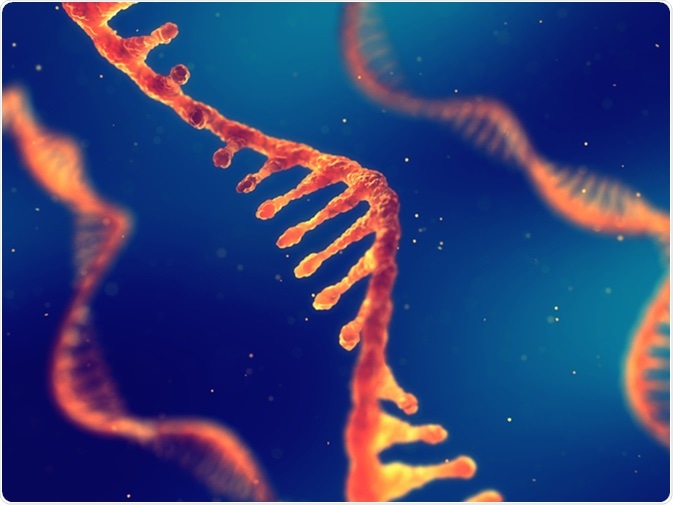
Single strand ribonucleic acid, RNA research 3d illustration. Image Credit: nobeastsofierce / Shutterstock
In vitro Evolution of Artificial Proteins and Enzymes
Methods to design artificial proteins in vitro include rational design and de novo selection. For these methods to succeed, a detailed knowledge of the domains of a protein and the way these fold is needed. However, lab evolution methods involve isolating artificial proteins and enzymes from libraries of established variants.
mRNA display method can be used select and evolve new proteins and enzymes from a large library of potential sequences. One method that has been recently proposed is to use mRNA display method to select for Diels-Alderase enzymes. Another method has also been proposed to employ this method to select for novel protein-ligand pairs by interaction-dependent reverse transcription. Also, generating tailor-made enzymes for various reactions is another potential area of application for mRNA display.
Self-Assembling Protein Microarrays
mRNA display technique generates mRNA-protein fusion sequences, which can be used for high throughput screening. These fusion sequences can code for MYC, HA11, or FLAG epitopes and subsequent incubation with DNA chips can lead to hybridization to the complementary regions. This can lead to self-directed assembly of the protein chip. These self-directed protein arrays can have the functionality of proteins that are displayed. Also, they are present in a uniform orientation and their detection limit is sub-attomole resolution.
Generating Non-Natural Libraries
The conventional methods to identify proteins are yeast two-hybrid and phage display. These two methods have an obligate in vivo step and they can only display the natural amino acid, of which there are 20. Including the amino acid alphabets can increase the chemical diversity and also improve the ligand diversity. The inclusion of un-natural amino acids can be done during translation or after translation to increase the diversity further.
During Translation
A recent method has shown that a suppressor tRNA strategy can be used to include unnatural amino acids, including biocytin (a lysine residue that is biotinylated), for the mRNA display. After the process of selection, the library can be enhanced for amber stop codon containing sequences. Using these methods can increase and enrich the diversity of the protein displayed, and this can further improve the chances of discovering novel molecules that have better specificity, stability or reactivity.
Post Translational Methods
In few recent studies, post translational methods for mRNA display have also been designed. In one such study, molecules that increased the inhibitory activity of penicillin towards penicillin binding protein 2a was selected from a library that contained pendant penicillin side chains. This strategy can be extended to other small molecule compounds and this can increase the diversity of display libraries of mRNA display technique.
Ligand Discovery
Having a larger library of potential sequences has several advantages: larger library can ease the discovery for higher affinity molecules, present greater diversity of molecules that have similar function, the large number of sequences that can be recovered can be further analyzed using other methods such as sequence covariation method. This method can be used to discover RNA-binding peptides, ATP aptamers, streptavidin aptamers, and TNF-α aptamers among others.
Using mRNA display, more than 100 RNA binding peptides have been discovered, and this represents one of the important functional test of mRNA display method. ATP aptamers were possibly the first proteins that bound to ATP. In a study, it was determined that one in 1012 molecules in mRNA library have ATP-binding ability.
Further Reading
Last Updated: Jul 20, 2023