Phase Focus Ltd (Phasefocus), the company that is revolutionizing microscopy and imaging with the Phasefocus Virtual Lens® technology, is pleased to report on a new paper published in Nature Scientific Reports describing their patented technology based on ptychography: "Ptychography - a label free, high-contrast imaging technique for live cells using quantitative phase information" is an open access paper written by a team of scientists from the Imaging and Cytometry Laboratory, part of the Bioscience Technology Facility in the Department of Biology at the University of York. The lead authors are Joanne Marrison and Peter O'Toole, the latter being head of the Laboratory.1

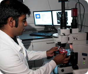
Dr Rakesh Suman uses the Phasefocus VL20 system at the University of York
Quoting from their summary, the authors describe the motivation for this work: "Cell imaging often relies on synthetic or genetic fluorescent labels, to provide contrast which can be far from ideal for imaging cells in their in vivo state. We report on the biological application of a label-free, high contrast microscopy technique known as ptychography, in which the image producing step is transferred from the microscope lens to a high-speed phase retrieval algorithm. We demonstrate that this technology is appropriate for label-free imaging of adherent cells and is particularly suitable for reporting cellular changes such as mitosis, apoptosis and cell differentiation. The high contrast, artefact-free, focus-free information rich images allow dividing cells to be distinguished from non-dividing cells by a greater than two-fold increase in cell contrast, and we demonstrate this technique is suitable for downstream automated cell segmentation and analysis."
The authors conclude that this has tremendous potential. "The system generates high quality images using inexpensive, low magnification objective lenses without the need for the addition of cytotoxic labels, a technical advance which has far reaching implications for cancer research and drug discovery. Not only are the images high in contrast, but they are also artefact free making them ideal for downstream segmentation and image analysis. The ability to focus post-acquisition allows all cells, even in an uneven field, to be brought into focus and not omitted from subsequent analysis. Time is also saved pre-acquisition as cells can be imaged directly in tissue culture plastic ware. We have shown here that this technique is suitable for cell cycle analysis, identifying cells entering and exiting mitosis and dissecting the phases of mitosis. Ptychography would be ideal for in-incubator assays, reporting over prolonged periods of time on cell proliferation and cell cycle stages in situ. These are only initial studies but clearly demonstrate the potential of label-free ptychographic imaging. With the technology rapidly developing, faster acquisition times, smart auto-focus, increased contrast and ease of use, and the ability to add the facility to other systems e.g. confocal microscopes, make the future opportunities diverse and very attractive."
Dr O'Toole describes ptychography as "one of the most important breakthroughs in imaging. It addresses many of the fundamental problems inherent in current microscopy techniques. Put simply, what you get is far more than what you see. Artefacts of sample preparation which may arise from labelling or use of markers are negated. I have been most impressed by Phasefocus and their ability to quickly respond to feedback and then to develop products to meet the needs of the user. I wonder where the technology will go? I feel the journey is just beginning and microscopy is nowhere near the end of the road with regards to development."
For more information on ptychography and its applications, please visit the Phasefocus web site: www.phasefocus.com.
Reference - the full article is available online at: http://www.nature.com/srep/2013/130806/srep02369/full/srep02369.html
1Ptychography - a label free, high-contrast imaging technique for live cells using quantitative phase information: Joanne Marrison, Lotta Räty, Poppy Marriott & Peter O'Toole. Scientific Reports 3, Article number: 2369 doi:10.1038/srep02369.