Phase Focus Ltd (Phasefocus), the company that is revolutionizing microscopy and imaging with the Phasefocus Virtual Lens® technology, reports on the Celldance competition results held at the recent annual ASCB meeting. KTP associate, Dr Rakesh Suman, was runner up with his video entitled "Cellular Heaven & Hell."
Time-lapse movies of a cellular "heaven and hell," a dividing crane fly sperm cell and the early development of muscle cells were recognized with the top three awards in the American Society for Cell Biology's Celldance "Really Useful" Cell Biology Video Contest for 2013. The annual Celldance awards were announced at the recent American Society of Cell Biology Annual Meeting in New Orleans. Simon Atkinson, chair of the ASCB Public Information Committee, which organizes Celldance, describes the awards as the "cellular Oscars." Says Atkinson, "Cell biology is the most visual of all the life sciences and, through Celldance, ASCB members are being encouraged to use microscopy videos as an outreach tool in education and on the Web."
Cellular Heaven and Hell - ASCB Runner Up!
Dr Rakesh Suman, a Phasefocus-sponsored Knowledge Transfer Partnership (KTP) Associate at the University of York shared the runners' up spot with his creation of Cellular Heaven and Hell, a time-lapse video obtained with the Phasefocus VL20 microscope, which uses ptychography to produce label-free, high-contrast, quantitative phase images. The healthy human bladder epithelial cells in "heaven" were imaged under normal physiological conditions every 10 minutes and are played back at seven frames per second. The human bladder epithelial cells in "hell" were treated with staurosporine to induce cell death, or apoptosis.

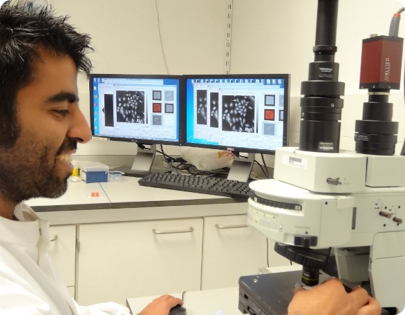
Dr Rakesh Suman, KTP Associate in the Imaging & Cytometry unit at the University of York using the Phasefocus VL20 system
Describing his work, Dr Suman says, "In the field of biological science, the imaging of key cellular events is usually visualized with the aid of fluorescent dyes. Although use of such dyes is commonplace and widely accepted, they do have their shortcomings as they can perturb the natural functionality of the cell."
"In my video, a novel technique known as ptychography was utilized to image apoptosis, a key cellular mechanism. I used the Phasefocus VL20 microscope to take high contrast and quantitative images of urothelial cells without using any labels or dyes. In this video, the cells in "heaven" are normal, healthy urothelial cells. Meanwhile the cells in "hell" were treated with the drug staurosporine to induce apoptosis. The microscope was able to clearly differentiate between these events."
"As the cells start to become apoptotic, they appear brighter in phase and adopt a more spherical morphology, up to the point where the cells rupture. Upon rupturing, the integrity of the cellular membrane is lost and there is no longer a separation between the intracellular and extracellular environment. Once again, this event is easily identifiable due to the quantitative nature of the images. A sharp and distinct drop in phase intensity is observed marking the end point of apoptosis."
This stunning video may be viewed on the Phasefocus YouTube channel: https://www.youtube.com/watch?v=krlMM-O0Ybc.
So, to summarise: the Phasefocus VL20 is shown to open up exciting avenues in biological imaging where cellular events and behaviours can be visualised and quantified without the use of any labels or dyes. For information on ptychography and its application for label-free, live cell imaging, please visit the Phasefocus web site: http://www.phasefocus.com/.