FEI (NASDAQ: FEIC) announced today its new Teneo VS™ scanning electron microscope (SEM), which offers a VolumeScope™ capability for life science applications. The Teneo platform tightly integrates FEI’s latest-generation SEM with VolumeScope, an in-chamber microtome and proprietary analytical software to provide fully-automated, large-volume reconstructions with dramatically improved z-axis resolution. The Teneo VS will be demonstrated at the International Microscopy Congress, to be held September 7-12, in Prague, Czech Republic. Initial shipments are planned for Q1 2015.

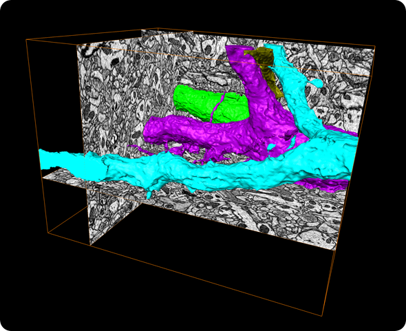
Volume reconstruction of mouse brain acquired with Teneo VS™. The block-face was imaged with the combination of Serial Block Face SEM and Multi-Energy Deconvolution SEM. 3D data visualization and reconstruction was done with Amira. Model depicting several axons (blue, purple and green). Isotropic pixels of 10 x 10 x 10 nm (x,y,z); Reconstructed volume 15.00 um x 12.9 um x 10.4 µm (1040 slices). Sample courtesy of P. Laserstein & P. Bastians, Helmstaedter Lab, MPI Frankfurt, Germany.
“The Teneo VS is a fully-integrated 3D volume imaging solution for life scientists,” said Peter Fruhstorfer, vice president and general manager of Life Sciences, FEI.
“Researchers in cellular, tissue, developmental and neurobiology, as well as toxicology and pharmacology, need to see nanoscale detail over relatively large sample volumes in order to understand functions and interactions within cells and tissues. The tight integration and extensive automation of the Teneo VS ensures fast, easy analysis, while the combination of our ThruSight™ and MAPS™ software deliver unprecedented resolution over large sample volumes.”
VolumeScope uses serial block face imaging (SBFI) to acquire a 3D volume from a block of tissue or cells. While SEMs can provide nanometer-scale lateral resolution in images of each slice, the axial resolution of the reconstructed model is normally limited by the physical thickness of the slices, typically 25 micrometers or more. The Teneo VS uses FEI’s ThruSight multi-energy deconvolution to resolve features at different depths within each slice, thus improving axial resolution.
The VolumeScope in-chamber ultra-microtome is fully-integrated with the Teneo VS operating and imaging software. Switching between volume imaging and normal SEM operation is fast and easy. MAPS software uses tiling and stitching to acquire high-resolution composite images that are much larger than a single field of view. MAPS also permits correlation with light microscope images for easy targeting of specific areas of interests. Reconstruction, visualization, analysis and presentation are handled by Amira™ software, which imports the image data directly from the microscope. The system also offers a low-vacuum water vapor option with dedicated detection that can reduce charging and provide further improvements in contrast and resolution on challenging samples.
For more information, please go to http://www.fei.com/teneo-for-life-sciences or visit the FEI booth (#6) at the International Microscopy Congress.