From algae to zebrafish, life under the microscope can be beautiful, surprising and mysterious. This week, amazing glimpses of the unseen universe earned top prizes in the 2014 Olympus BioScapes Digital Imaging Competition®, the world’s foremost forum for showcasing microscope images of life science subjects.
First Prize went to an extraordinary movie of a fruit fly developing, captured by a team including William Lemon, Fernando Amat and Philipp Keller of the Howard Hughes Medical Institute (HHMI) Janelia Research Campus in Ashburn, Va. In the fascinating video, a trembling ball of cells turns into a fully developed fly larva that actually starts to crawl off screen by the movie’s end.
It was selected from among nearly 2500 entries to earn $5,000 worth of Olympus equipment. Significantly, the winning team is donating its First Prize to the Children’s Science Center, a new museum that will be opening in Northern Virginia in 2015. All BioScapes award-winning images may be viewed online at OlympusBioScapes.com.

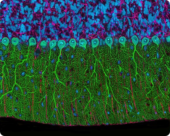
Thomas Deerinck, Rat cerebellum. 2nd Prize, 2014 Olympus BioScapes Digital Imaging Competition
Celebrating its 11th year, the Olympus BioScapes Competition is the world’s premier platform for honoring images and movies of human, plant and animal subjects as captured through light microscopes. Images of any life science subject are eligible. Entries are judged based on the science they depict, their beauty or impact, and the technical expertise involved in capturing them. Entrants can use any brand of light microscope. In addition to the Top 10 award-winning recipients, 62 Honorable Mentions and one Technical Merit Award were distributed this year. Nine movies were among the winners.
The 2014 winning entries reflect the latest advances in neuroscience and developmental biology as documented by researchers. The First Prize movie is a good example. High-speed videos like this allow researchers, almost for the first time, to follow the fate of individual embryonic cells from shortly after fertilization to the time the fly larva hatches; the cells divide, migrate, diversify and eventually become the varied organs and systems of the fly. Understanding exactly how an animal develops and how different cells develop separate functions can lead to a better understanding of life processes, and ultimately may contribute to disease research. The movie, captured using a custom-built simultaneous multi-view light sheet microscope, includes more than two million images in the complete data set; its true potential is extracted by using complex mathematical models to learn the fate of every cell.
Second Prize was a brilliant photo of a rat cerebellum, part of the brain, captured by Thomas Deerinck of the University of California San Diego’s National Center for Microscopy and Imaging Research. The image, captured using multiphoton imaging, was captured at 300x.
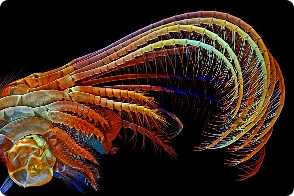
Other images offer additional eye-opening glimpses of life on a microscopic scale as captured by scientists, hobbyists and students. Igor Siwanowicz, another researcher at HHMI Janelia Research Campus, earned Third Prize for his colorful confocal image of the parts of a barnacle that sweep food into the barnacle’s shell for consumption. Fourth prize was an amusing image of two weevils staged and captured by Csaba Pintér of Keszthely, Hungary.

Igor Siwanowicz, Barnacle appendages. 3rd Prize, 2014 Olympus BioScapes Digital Imaging Competition
This year’s honored images come from 14 states of the U.S. and 21 other nations: Belgium, Canada, Chile, Denmark, England, France, Germany, Hungary, Iran, Ireland, Italy, Japan, The Netherlands, Norway, Panama, Poland, Russian Federation, Singapore, Spain, Sweden and the United Arab Emirates. Competition participants hailed from 68 nations, making this year’s competition among the most international to date.
One of the most interesting images this year was the Ninth Prize image of the gears of a green coneheaded planthopper (Acanalonia conica) nymph. The insects are accomplished jumpers, able to accelerate at staggering 500 times the force of gravity. To synchronize the movement of their hind legs, their trochanters are coupled with a pair of cogs. Images such as this one help demonstrate that gears, which until recently were thought to be a human invention, exist in the natural world. The confocal image was captured by Igor Siwanowicz.
The other Top 10 prizewinners: Fifth Prize, a confocal image of rat brain cerebral cortex, was captured by Madelyn May, of Hano, N.H. David Johnston, of Southampton General Hospital, Southampton, UK, earned Sixth Prize for a captivating confocal image of a worm larva from plankton collected off the coast of England. Oleksandr Holovachov, of Ekuddsvagen, Sweden, took Seventh Prize for a fluorescence photo of a butter daisy. Matthew S. Lehnert and Ashley L. Lash, Kent State University at Stark, North Canton, Ohio, captured Eighth Prize for a confocal image of the proboscis (mouthparts) of a vampire moth. Tenth Prize this year went to a fascinating video of the neural activity in an entire living zebrafish brain captured by Philipp Keller, Fernando Amat and Misha Ahrens of HHMI Janelia Research Campus, Ashburn, Va. The video shows fast 3D recordings of the entire larval brain (about 100,000 neurons) and depicts, for the first time, an almost exhaustive view of single-neuron activity in the brain of a vertebrate.
“For 11 years, Olympus has sponsored this competition to shed light on the importance of research and draw attention to the amazing intersection of science and art,” said Hidenao Tsuchiya, Chairman of Olympus Scientific Solutions Americas, part of Olympus Corporation.
Olympus BioScapes movies and images have spurred public interest in and support of microscopy, drawn attention to the vital work that goes on in laboratories worldwide, and inspired young people to seek careers in science.
A selection of the 2014 winning and Honorable Mention images and videos will be displayed in a museum tour that will travel the U.S. over the coming year. The 2014 BioScapes tour is sponsored by Olympus in cooperation with Scientific American. Other exhibits of winning BioScapes images also will be shown across the U.S., Mexico, Latin America and Canada in 2015.
Olympus selects outstanding authorities in microscope imaging as judges for each year’s competition. This year’s BioScapes panel of judges included the eminent Robert E. Campbell, Ph.D., University of Alberta, Edmonton, Alta., Canada; Catherine Galbraith, Ph.D., Oregon Health and Science University Knight Cancer Institute, Portland Ore.; Douglas Murphy, Ph.D., HHMI Janelia Research Campus, Ashburn, Va.; and Alison North, Ph.D., The Rockefeller University, N.Y. The judging took place under the direction of Michael W. Davidson, The Florida State University, Tallahassee, Fla., and his staff.
The Olympus BioScapes Competition was founded to highlight the amazing stories coming out of laboratories around the world, and to inspire excellence in science and scientific photography. Entrants can submit up to five still images, image sequences, or movies of life science subjects captured at any magnification using a compound light microscope. The judges make their decisions without participant or brand information. To view the BioScapes gallery of winners and Honorable Mentions, visit www.OlympusBioScapes.com.
About Olympus
Olympus Corporation is an international precision technology leader operating in life science, industrial, medical, academic and consumer markets, specializing in optics, electronics, and precision engineering. As a subsidiary of Olympus Corporation, Olympus Scientific Solutions Americas is an integral part of the global Olympus network with specific responsibility for the sales and marketing of life science and industrial instrumentation in the Americas. The company’s core product lineup comprises clinical, educational, and research microscopes, nondestructive testing equipment, and analytical instruments all designed with the unwavering commitment to enhancing people’s lives every day and contributing to the safety, security, quality, and productivity of society. For more information, visit www.olympusamerica.com