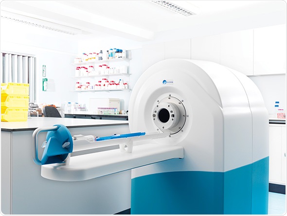
Manchester University has chosen MR Solutions’ 3T MRI preclinical, cryogen free imaging solution for superior soft tissue contrast and molecular imaging research at the Wolfson Molecular Imaging Centre. The scanner will be used in their Regenerative Medicine research programme which aims to develop a clearer understanding of the potential risks and hazards of therapies and to develop new methodologies to assess their risk. The aim of this research is to gain faster access to new medicines for patients.

This new state of the art preclinical imaging system was developed, supplied and installed by MR Solutions of Guildford, Surrey and supported by the UK Regenerative Medicine Platform with a grant for equipment to the Stem Cell Safety Hub.
This is a multi-institutional collaboration led by Liverpool University with Manchester, UCL and Edinburgh universities: see http://www.ukrmp.org.uk/hubs/safety/. The UK Regenerative Medicine Platform (UKRMP) is a £25M initiative that is addressing the key translational challenges of regenerative medicine. Established in 2013 by BBSRC, EPSRC and MRC, the UKRMP provides critical mass of academic expertise, innovation and knowledge, connected to commercial and clinical end-users.
Professor Steve Williams is the Centre Lead for Imaging Sciences and explained:
We wanted a system with a small footprint which could be located alongside other imaging equipment in our laboratory. The cryogen-free 3T preclinical system has a superconducting, dry magnet with almost no fringe field allowing safe use within our existing laboratory
MR Solutions which developed the ground-breaking dry magnet technology is the only company across the globe to have supplied these scanners to laboratories across the world. The 3T system does away with the need for liquid helium, which reduces running costs and there is no requirement for building modification works. It can be placed near other sensitive equipment as it has a stray magnetic field of only a few centimetres.
MR Solutions has also pioneered multi-modality imaging which dispenses with the need for separate systems. PET-MRI or SPECT-MRI imaging either for independent acquisition, sequential acquisition, or simultaneous acquisition can be facilitated. Optical and CT imaging capabilities are also possible.
Dr David Taylor, of MR Solutions said:
Multi-modality imaging will become the norm for the study of anatomy, bio-distribution, efficacy, safety and kinetics within the same anatomical context. It provides much better results all within a scanner which can sit on a desk